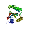
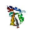
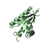
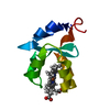
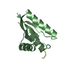
- PDB-3o1f: P1 crystal form of E. coli ClpS at 1.4 A resolution -
+
Open data
ID or keywords:
Loading...
-
Basic information
Entry
Database: PDB / ID: 3o1f
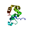
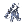
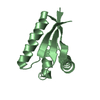
Title
P1 crystal form of E. coli ClpS at 1.4 A resolution
Components
(ATP-dependent Clp protease adapter protein clpS) x 2
Keywords
HYDROLASE / adaptor
Function / homology
Function and homology information
molecular function inhibitor activity / protein catabolic process / peptidase activity / response to heat / protein-folding chaperone binding / proteolysis Similarity search - Function
Ribosomal protein L7/L12, C-terminal domain/Adaptor protein ClpS / ATP-dependent Clp protease adaptor protein ClpS / Adaptor protein ClpS, core / ATP-dependent Clp protease adaptor protein ClpS / Ribosomal Protein L30; Chain: A, / Ribosomal protein L7/L12, C-terminal/adaptor protein ClpS-like / 2-Layer Sandwich / Alpha Beta Similarity search - Domain/homology
ATP-dependent Clp protease adapter protein ClpS / ATP-dependent Clp protease adapter protein ClpS Similarity search - Component
Biological species
Escherichia coli (E. coli)
Method
X-RAY DIFFRACTION / MOLECULAR REPLACEMENT / Resolution: 1.4 Å
In the structure databanks used in Yorodumi, some data are registered as the other names, "COVID-19 virus" and "2019-nCoV". Here are the details of the virus and the list of structure data.
Jan 31, 2019. EMDB accession codes are about to change! (news from PDBe EMDB page)
EMDB accession codes are about to change! (news from PDBe EMDB page)
The allocation of 4 digits for EMDB accession codes will soon come to an end. Whilst these codes will remain in use, new EMDB accession codes will include an additional digit and will expand incrementally as the available range of codes is exhausted. The current 4-digit format prefixed with “EMD-” (i.e. EMD-XXXX) will advance to a 5-digit format (i.e. EMD-XXXXX), and so on. It is currently estimated that the 4-digit codes will be depleted around Spring 2019, at which point the 5-digit format will come into force.
The EM Navigator/Yorodumi systems omit the EMD- prefix.
Related info.:Q: What is EMD? / ID/Accession-code notation in Yorodumi/EM Navigator
Yorodumi is a browser for structure data from EMDB, PDB, SASBDB, etc.
This page is also the successor to EM Navigator detail page, and also detail information page/front-end page for Omokage search.
The word "yorodu" (or yorozu) is an old Japanese word meaning "ten thousand". "mi" (miru) is to see.
Related info.:EMDB / PDB / SASBDB / Comparison of 3 databanks / Yorodumi Search / Aug 31, 2016. New EM Navigator & Yorodumi / Yorodumi Papers / Jmol/JSmol / Function and homology information / Changes in new EM Navigator and Yorodumi
 Movie
Movie Controller


 Open data
 Basic information
 Components
 Keywords
 Function and homology information
 X-RAY DIFFRACTION /
  Authors
 Citation
 Structure visualization
 Downloads & links
 Other downloads


 PDBj
PDBj

 Assembly
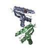
 Mass: 18.015 Da / Num. of mol.: 402 / Source method: isolated from a natural source / Formula: H2O
 Sample preparation
 Processing