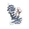
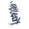
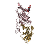
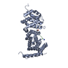
Entry Database : PDB / ID : 3nmwTitle Crystal structure of armadillo repeats domain of APC APC variant protein Keywords / / Function / homology Function Domain/homology Component
/ / / / / / / / / / / / / / / / / / / / / / / / / / / / / / / / / / / / / / / / / / / / / / / / / / / / / / / / / / / / / / / / / / / / / / / / / / / / / / / / / / / / / / / / / / / / / / / / / / / / / / / / / / / / / / / / / / / / / / / / / / / / / / / Biological species Homo sapiens (human)Method / / / Resolution : 1.6 Å Authors Zhang, Z. / Chen, L. / Gao, L. / Lin, K. / Wu, G. Journal : Cell Res. / Year : 2012Title : Structural basis for the recognition of Asef by adenomatous polyposis coli.Authors : Zhang, Z. / Chen, L. / Gao, L. / Lin, K. / Zhu, L. / Lu, Y. / Shi, X. / Gao, Y. / Zhou, J. / Xu, P. / Zhang, J. / Wu, G. History Deposition Jun 22, 2010 Deposition site / Processing site Revision 1.0 Jul 6, 2011 Provider / Type Revision 1.1 Jul 13, 2011 Group Revision 1.2 Dec 21, 2016 Group Revision 1.3 Mar 15, 2017 Group Revision 1.4 Dec 25, 2019 Group / Database references / Derived calculationsCategory citation / pdbx_unobs_or_zero_occ_atoms ... citation / pdbx_unobs_or_zero_occ_atoms / struct_conn / struct_ref_seq_dif Item _citation.pdbx_database_id_PubMed / _citation.title ... _citation.pdbx_database_id_PubMed / _citation.title / _struct_conn.pdbx_leaving_atom_flag / _struct_ref_seq_dif.details Revision 1.5 Dec 27, 2023 Group Advisory / Data collection ... Advisory / Data collection / Database references / Derived calculations / Refinement description Category chem_comp_atom / chem_comp_bond ... chem_comp_atom / chem_comp_bond / database_2 / pdbx_unobs_or_zero_occ_atoms / struct_ncs_dom_lim / struct_site Item _database_2.pdbx_DOI / _database_2.pdbx_database_accession ... _database_2.pdbx_DOI / _database_2.pdbx_database_accession / _struct_ncs_dom_lim.beg_auth_comp_id / _struct_ncs_dom_lim.beg_label_asym_id / _struct_ncs_dom_lim.beg_label_comp_id / _struct_ncs_dom_lim.beg_label_seq_id / _struct_ncs_dom_lim.end_auth_comp_id / _struct_ncs_dom_lim.end_label_asym_id / _struct_ncs_dom_lim.end_label_comp_id / _struct_ncs_dom_lim.end_label_seq_id / _struct_site.pdbx_auth_asym_id / _struct_site.pdbx_auth_comp_id / _struct_site.pdbx_auth_seq_id Revision 1.6 Oct 16, 2024 Group / Category / pdbx_modification_feature
Show all Show less
 Movie
Movie Controller


 Open data
 Basic information
 Components
 Keywords
 Function and homology information
 Homo sapiens (human)
 X-RAY DIFFRACTION /
  Authors
 Citation
 Structure visualization
 Downloads & links
 Other downloads
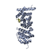


 PDBj
PDBj


 Assembly
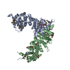


 Mass: 96.063 Da / Num. of mol.: 2 / Source method: obtained synthetically / Formula: SO4
 Mass: 18.015 Da / Num. of mol.: 680 / Source method: isolated from a natural source / Formula: H2O
 Sample preparation
 / Beamline: BL17U / Wavelength: 0.97916 Å
 Processing