| Entry | Database: PDB / ID: 4yje
|
|---|
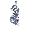
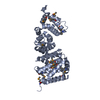
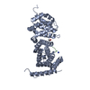
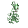
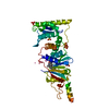
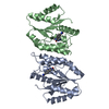
| Title | Crystal structure of APC-ARM in complexed with Amer1-A1 |
|---|
 Components Components | - APC membrane recruitment protein 1
- Adenomatous polyposis coli protein
|
|---|
 Keywords Keywords | CELL ADHESION/PROTEIN BINDING / ARMADILLO-LIGAND COMPLEX / CELL ADHESION-PROTEIN BINDING COMPLEX |
|---|
| Function / homology |  Function and homology information Function and homology information
APC truncation mutants are not K63 polyubiquitinated / beta-catenin destruction complex binding / negative regulation of cell cycle G1/S phase transition / negative regulation of cyclin-dependent protein serine/threonine kinase activity / gamma-catenin binding / regulation of attachment of spindle microtubules to kinetochore / pattern specification process / positive regulation of pseudopodium assembly / positive regulation of protein localization to centrosome / negative regulation of microtubule depolymerization ...APC truncation mutants are not K63 polyubiquitinated / beta-catenin destruction complex binding / negative regulation of cell cycle G1/S phase transition / negative regulation of cyclin-dependent protein serine/threonine kinase activity / gamma-catenin binding / regulation of attachment of spindle microtubules to kinetochore / pattern specification process / positive regulation of pseudopodium assembly / positive regulation of protein localization to centrosome / negative regulation of microtubule depolymerization / bicellular tight junction assembly / heart valve development / beta-catenin destruction complex / catenin complex / APC truncation mutants have impaired AXIN binding / AXIN missense mutants destabilize the destruction complex / Truncations of AMER1 destabilize the destruction complex / microtubule plus-end binding / protein kinase regulator activity / Beta-catenin phosphorylation cascade / Signaling by GSK3beta mutants / CTNNB1 S33 mutants aren't phosphorylated / CTNNB1 S37 mutants aren't phosphorylated / CTNNB1 S45 mutants aren't phosphorylated / CTNNB1 T41 mutants aren't phosphorylated / cell fate specification / Wnt signalosome / regulation of microtubule-based process / Disassembly of the destruction complex and recruitment of AXIN to the membrane / regulation of canonical Wnt signaling pathway / endocardial cushion morphogenesis / Apoptotic cleavage of cellular proteins / mitotic spindle assembly checkpoint signaling / negative regulation of G1/S transition of mitotic cell cycle / regulation of microtubule-based movement / mitotic cytokinesis / dynein complex binding / lateral plasma membrane / bicellular tight junction / phosphatidylinositol-4,5-bisphosphate binding / positive regulation of protein ubiquitination / adherens junction / Deactivation of the beta-catenin transactivating complex / negative regulation of canonical Wnt signaling pathway / beta-catenin binding / kinetochore / Degradation of beta-catenin by the destruction complex / Wnt signaling pathway / ruffle membrane / positive regulation of protein catabolic process / positive regulation of canonical Wnt signaling pathway / insulin receptor signaling pathway / Ovarian tumor domain proteases / KEAP1-NFE2L2 pathway / nervous system development / cell migration / lamellipodium / positive regulation of cold-induced thermogenesis / Neddylation / protein-containing complex assembly / microtubule binding / microtubule / proteasome-mediated ubiquitin-dependent protein catabolic process / cell adhesion / nuclear body / positive regulation of cell migration / positive regulation of apoptotic process / negative regulation of cell population proliferation / centrosome / ubiquitin protein ligase binding / DNA damage response / protein kinase binding / perinuclear region of cytoplasm / Golgi apparatus / nucleoplasm / nucleus / plasma membrane / cytosol / cytoplasmSimilarity search - Function APC membrane recruitment protein / APC membrane recruitment protein / Adenomatous polyposis coli protein repeat / SAMP / EB-1 binding / Adenomatous polyposis coli protein basic domain / Adenomatous polyposis coli protein, 15 residue repeat / Adenomatous polyposis coli (APC) family / Adenomatous polyposis coli protein / Adenomatous polyposis coli, N-terminal dimerisation domain ...APC membrane recruitment protein / APC membrane recruitment protein / Adenomatous polyposis coli protein repeat / SAMP / EB-1 binding / Adenomatous polyposis coli protein basic domain / Adenomatous polyposis coli protein, 15 residue repeat / Adenomatous polyposis coli (APC) family / Adenomatous polyposis coli protein / Adenomatous polyposis coli, N-terminal dimerisation domain / APC, N-terminal coiled-coil domain superfamily / Adenomatous polyposis coli (APC) repeat / APC repeat / SAMP Motif / EB-1 Binding Domain / APC basic domain / APC 15 residue motif / Adenomatous polyposis coli tumour suppressor protein / Armadillo-associated region on APC / Unstructured region on APC between 1st and 2nd catenin-bdg motifs / Unstructured region on APC between 1st two creatine-rich regions / Unstructured region on APC between APC_crr and SAMP / Unstructured region on APC between SAMP and APC_crr / Unstructured region on APC between APC_crr regions 5 and 6 / Coiled-coil N-terminus of APC, dimerisation domain / Adenomatous polyposis coli (APC) repeat / Armadillo/plakoglobin ARM repeat profile. / Armadillo/beta-catenin-like repeat / Armadillo/beta-catenin-like repeats / Armadillo / Leucine-rich Repeat Variant / Leucine-rich Repeat Variant / Alpha Horseshoe / Armadillo-like helical / Armadillo-type fold / Mainly AlphaSimilarity search - Domain/homology |
|---|
| Biological species |  Homo sapiens (human) Homo sapiens (human) |
|---|
| Method |  X-RAY DIFFRACTION / SYNCHROTRON / MOLECULAR REPLACEMENT / Resolution: 1.9 Å X-RAY DIFFRACTION / SYNCHROTRON / MOLECULAR REPLACEMENT / Resolution: 1.9 Å |
|---|
 Authors Authors | Zhang, Z. / Xiao, Y. / Wu, G. |
|---|
 Citation Citation | Journal: Cell Discov / Year: 2015
Title: Structures of the APC-ARM domain in complexes with discrete Amer1/WTX fragments reveal that it uses a consensus mode to recognize its binding partners
Authors: Zhang, Z. / Akyildiz, S. / Xiao, Y. / Gai, Z. / An, Y. / Behrens, J. / Wu, G. |
|---|
| History | | Deposition | Mar 3, 2015 | Deposition site: RCSB / Processing site: PDBJ |
|---|
| Revision 1.0 | Mar 9, 2016 | Provider: repository / Type: Initial release |
|---|
| Revision 1.1 | Aug 17, 2016 | Group: Database references |
|---|
| Revision 1.2 | Dec 21, 2016 | Group: Structure summary |
|---|
| Revision 1.3 | Nov 8, 2023 | Group: Data collection / Database references / Refinement description
Category: chem_comp_atom / chem_comp_bond ...chem_comp_atom / chem_comp_bond / database_2 / pdbx_initial_refinement_model
Item: _database_2.pdbx_DOI / _database_2.pdbx_database_accession |
|---|
|
|---|
 Movie
Movie Controller


 Open data
 Basic information
 Structure visualization
 Downloads & links
 Other downloads
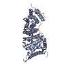


 PDBj
PDBj


 Assembly
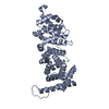

 Mass: 18.015 Da / Num. of mol.: 265 / Source method: isolated from a natural source / Formula: H2O
 Sample preparation
 / Beamline: BL17U / Wavelength: 0.97935 Å
 Processing