 Movie
Movie Controller


+ Open data

- Basic information
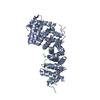
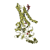
| Entry | Database: PDB / ID: 4yjl | ||||||
|---|---|---|---|---|---|---|---|
| Title | Crystal structure of APC-ARM in complexed with Amer1-A2 | ||||||
 Components Components |
| ||||||
 Keywords Keywords | CELL ADHESION/PROTEIN BINDING / ARMADILLO-LIGAND COMPLEX / CELL ADHESION-PROTEIN BINDING COMPLEX | ||||||
| Function / homology |  Function and homology information Function and homology informationAPC truncation mutants are not K63 polyubiquitinated / beta-catenin destruction complex binding / negative regulation of cell cycle G1/S phase transition / negative regulation of cyclin-dependent protein serine/threonine kinase activity / gamma-catenin binding / regulation of attachment of spindle microtubules to kinetochore / positive regulation of pseudopodium assembly / positive regulation of protein localization to centrosome / bicellular tight junction assembly / negative regulation of microtubule depolymerization ...APC truncation mutants are not K63 polyubiquitinated / beta-catenin destruction complex binding / negative regulation of cell cycle G1/S phase transition / negative regulation of cyclin-dependent protein serine/threonine kinase activity / gamma-catenin binding / regulation of attachment of spindle microtubules to kinetochore / positive regulation of pseudopodium assembly / positive regulation of protein localization to centrosome / bicellular tight junction assembly / negative regulation of microtubule depolymerization / pattern specification process / heart valve development / beta-catenin destruction complex / catenin complex / APC truncation mutants have impaired AXIN binding / AXIN missense mutants destabilize the destruction complex / Truncations of AMER1 destabilize the destruction complex / microtubule plus-end binding / protein kinase regulator activity / Beta-catenin phosphorylation cascade / Signaling by GSK3beta mutants / CTNNB1 S33 mutants aren't phosphorylated / CTNNB1 S37 mutants aren't phosphorylated / CTNNB1 S45 mutants aren't phosphorylated / CTNNB1 T41 mutants aren't phosphorylated / Wnt signalosome / regulation of microtubule-based process / cell fate specification / Disassembly of the destruction complex and recruitment of AXIN to the membrane / regulation of canonical Wnt signaling pathway / endocardial cushion morphogenesis / Apoptotic cleavage of cellular proteins / mitotic spindle assembly checkpoint signaling / negative regulation of G1/S transition of mitotic cell cycle / regulation of microtubule-based movement / mitotic cytokinesis / dynein complex binding / lateral plasma membrane / bicellular tight junction / phosphatidylinositol-4,5-bisphosphate binding / positive regulation of protein ubiquitination / adherens junction / Deactivation of the beta-catenin transactivating complex / negative regulation of canonical Wnt signaling pathway / kinetochore / beta-catenin binding / Degradation of beta-catenin by the destruction complex / ruffle membrane / Wnt signaling pathway / positive regulation of protein catabolic process / insulin receptor signaling pathway / Ovarian tumor domain proteases / KEAP1-NFE2L2 pathway / positive regulation of canonical Wnt signaling pathway / nervous system development / cell migration / lamellipodium / positive regulation of cold-induced thermogenesis / Neddylation / protein-containing complex assembly / microtubule binding / microtubule / proteasome-mediated ubiquitin-dependent protein catabolic process / cell adhesion / nuclear body / positive regulation of cell migration / positive regulation of apoptotic process / negative regulation of cell population proliferation / DNA damage response / ubiquitin protein ligase binding / centrosome / protein kinase binding / perinuclear region of cytoplasm / Golgi apparatus / nucleoplasm / nucleus / plasma membrane / cytoplasm / cytosol Similarity search - Function | ||||||
| Biological species |  Homo sapiens (human) Homo sapiens (human) | ||||||
| Method |  X-RAY DIFFRACTION / SYNCHROTRON / MOLECULAR REPLACEMENT / Resolution: 2.1 Å X-RAY DIFFRACTION / SYNCHROTRON / MOLECULAR REPLACEMENT / Resolution: 2.1 Å | ||||||
 Authors Authors | Zhang, Z. / Xiao, Y. / Wu, G. | ||||||
 Citation Citation | Journal: Cell Discov / Year: 2015 Title: Structures of the APC-ARM domain in complexes with discrete Amer1/WTX fragments reveal that it uses a consensus mode to recognize its binding partners Authors: Zhang, Z. / Akyildiz, S. / Xiao, Y. / Gai, Z. / An, Y. / Behrens, J. / Wu, G. | ||||||
| History |
|
- Structure visualization
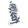
| Structure viewer | Molecule: MolmilJmol/JSmol |
|---|
- Downloads & links
-Download
| PDBx/mmCIF format | 4yjl.cif.gz | 462.5 KB | Display | PDBx/mmCIF format |
|---|---|---|---|---|
| PDB format | pdb4yjl.ent.gz | 378 KB | Display | PDB format |
| PDBx/mmJSON format | 4yjl.json.gz | Tree view | PDBx/mmJSON format | |
| Others |  Other downloads Other downloads |
-Validation report
| Arichive directory | https://data.pdbj.org/pub/pdb/validation_reports/yj/4yjlftp://data.pdbj.org/pub/pdb/validation_reports/yj/4yjl | HTTPS FTP |
|---|
-Related structure data
| Related structure data |  4yjeC 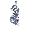 4yk6C 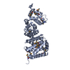 3nmwS C: citing same article ( S: Starting model for refinement |
|---|---|
| Similar structure data |


-Links
 PDBj
PDBj


- Assembly
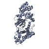
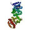
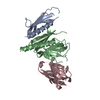
| Deposited unit | 
| ||||||||
|---|---|---|---|---|---|---|---|---|---|
| 1 | 
| ||||||||
| 2 | 
| ||||||||
| 3 | 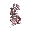
| ||||||||
| 4 | 
| ||||||||
| 5 | 
| ||||||||
| 6 | 
| ||||||||
| Unit cell |
|
-Components
| #1: Protein | Mass: 39268.246 Da / Num. of mol.: 6 / Fragment: ARM DOMAIN, UNP RESIDUES 407-751 Source method: isolated from a genetically manipulated source Source: (gene. exp.) Homo sapiens (human) / Gene: APC / Plasmid: PET28A / Production host:  #2: Protein/peptide | Mass: 1520.578 Da / Num. of mol.: 6 / Fragment: UNP RESIDUES 496-508 / Source method: obtained synthetically / Details: This sequence occurs naturally in humans. / Source: (synth.) Homo sapiens (human) / References: UniProt: Q5JTC6#3: Chemical | ChemComp-EDO /   Mass: 62.068 Da / Num. of mol.: 37 / Source method: obtained synthetically / Formula: C2H6O2 Mass: 62.068 Da / Num. of mol.: 37 / Source method: obtained synthetically / Formula: C2H6O2#4: Water | ChemComp-HOH / |  Mass: 18.015 Da / Num. of mol.: 2397 / Source method: isolated from a natural source / Formula: H2O Mass: 18.015 Da / Num. of mol.: 2397 / Source method: isolated from a natural source / Formula: H2O |
|---|
-Experimental details
-Experiment
| Experiment | Method: X-RAY DIFFRACTION / Number of used crystals: 1 |
|---|
- Sample preparation
| Crystal | Density Matthews: 6.18 Å3/Da / Density % sol: 80.11 % |
|---|---|
| Crystal grow | Temperature: 287 K / Method: vapor diffusion, hanging drop / pH: 6.2 / Details: 0.2M NACL, 10% PEG 8000 |
-Data collection
| Diffraction | Mean temperature: 100 K |
|---|---|
| Diffraction source | Source: SYNCHROTRON / Site: SSRF  / Beamline: BL17U / Wavelength: 0.97935 Å / Beamline: BL17U / Wavelength: 0.97935 Å |
| Detector | Type: ADSC QUANTUM 315r / Detector: CCD / Date: Nov 8, 2011 |
| Radiation | Protocol: SINGLE WAVELENGTH / Monochromatic (M) / Laue (L): M / Scattering type: x-ray |
| Radiation wavelength | Wavelength: 0.97935 Å / Relative weight: 1 |
| Reflection | Resolution: 2.1→50 Å / Num. obs: 335882 / % possible obs: 98.2 % / Redundancy: 3.9 % / Rmerge(I) obs: 0.103 / Net I/σ(I): 7.7 |
| Reflection shell | Resolution: 2.1→2.18 Å / Redundancy: 3.9 % / Rmerge(I) obs: 0.527 / % possible all: 97.3 |
- Processing
| Software |
| ||||||||||||||||||||||||||||||||||||||||||||||||||||||||||||||||||||||||||||||||||||||||||||||||||||||||||||||||||||||||||||||||||||||||||||||||||||||||||||||||||||||||||||||||||||||
|---|---|---|---|---|---|---|---|---|---|---|---|---|---|---|---|---|---|---|---|---|---|---|---|---|---|---|---|---|---|---|---|---|---|---|---|---|---|---|---|---|---|---|---|---|---|---|---|---|---|---|---|---|---|---|---|---|---|---|---|---|---|---|---|---|---|---|---|---|---|---|---|---|---|---|---|---|---|---|---|---|---|---|---|---|---|---|---|---|---|---|---|---|---|---|---|---|---|---|---|---|---|---|---|---|---|---|---|---|---|---|---|---|---|---|---|---|---|---|---|---|---|---|---|---|---|---|---|---|---|---|---|---|---|---|---|---|---|---|---|---|---|---|---|---|---|---|---|---|---|---|---|---|---|---|---|---|---|---|---|---|---|---|---|---|---|---|---|---|---|---|---|---|---|---|---|---|---|---|---|---|---|---|---|
| Refinement | Method to determine structure: MOLECULAR REPLACEMENT Starting model: 3NMW Resolution: 2.1→49.34 Å / Cor.coef. Fo:Fc: 0.932 / Cor.coef. Fo:Fc free: 0.919 / Cross valid method: THROUGHOUT / σ(F): 0 / ESU R: 0.125 / ESU R Free: 0.118 / Stereochemistry target values: MAXIMUM LIKELIHOOD / Details: HYDROGENS HAVE BEEN USED IF PRESENT IN
| ||||||||||||||||||||||||||||||||||||||||||||||||||||||||||||||||||||||||||||||||||||||||||||||||||||||||||||||||||||||||||||||||||||||||||||||||||||||||||||||||||||||||||||||||||||||
| Solvent computation | Ion probe radii: 0.8 Å / Shrinkage radii: 0.8 Å / VDW probe radii: 1.2 Å / Solvent model: MASK | ||||||||||||||||||||||||||||||||||||||||||||||||||||||||||||||||||||||||||||||||||||||||||||||||||||||||||||||||||||||||||||||||||||||||||||||||||||||||||||||||||||||||||||||||||||||
| Displacement parameters | Biso mean: 27.01 Å2
| ||||||||||||||||||||||||||||||||||||||||||||||||||||||||||||||||||||||||||||||||||||||||||||||||||||||||||||||||||||||||||||||||||||||||||||||||||||||||||||||||||||||||||||||||||||||
| Refinement step | Cycle: LAST / Resolution: 2.1→49.34 Å
| ||||||||||||||||||||||||||||||||||||||||||||||||||||||||||||||||||||||||||||||||||||||||||||||||||||||||||||||||||||||||||||||||||||||||||||||||||||||||||||||||||||||||||||||||||||||
| Refine LS restraints |
|