 Movie
Movie Controller


+ Open data

- Basic information
| Entry | Database: PDB / ID: 3ndf | ||||||
|---|---|---|---|---|---|---|---|
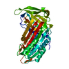
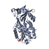
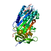
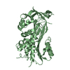
| Title | Cleaved antitrypsin with P8-P6 Asp | ||||||
 Components Components | (Alpha-1-antitrypsin) x 2 | ||||||
 Keywords Keywords | HYDROLASE INHIBITOR / serpin | ||||||
| Function / homology |  Function and homology information Function and homology informationCargo concentration in the ER / COPII-coated ER to Golgi transport vesicle / COPII-mediated vesicle transport / endoplasmic reticulum-Golgi intermediate compartment membrane / platelet alpha granule lumen / acute-phase response / Post-translational protein phosphorylation / serine-type endopeptidase inhibitor activity / Regulation of Insulin-like Growth Factor (IGF) transport and uptake by Insulin-like Growth Factor Binding Proteins (IGFBPs) / blood coagulation ...Cargo concentration in the ER / COPII-coated ER to Golgi transport vesicle / COPII-mediated vesicle transport / endoplasmic reticulum-Golgi intermediate compartment membrane / platelet alpha granule lumen / acute-phase response / Post-translational protein phosphorylation / serine-type endopeptidase inhibitor activity / Regulation of Insulin-like Growth Factor (IGF) transport and uptake by Insulin-like Growth Factor Binding Proteins (IGFBPs) / blood coagulation / Platelet degranulation / extracellular matrix / protease binding / ficolin-1-rich granule lumen / endoplasmic reticulum lumen / Neutrophil degranulation / Golgi apparatus / endoplasmic reticulum / : / extracellular exosome / extracellular region / identical protein binding Similarity search - Function | ||||||
| Biological species |  Homo sapiens (human) Homo sapiens (human) | ||||||
| Method |  X-RAY DIFFRACTION / MOLECULAR REPLACEMENT / Resolution: 2.7 Å X-RAY DIFFRACTION / MOLECULAR REPLACEMENT / Resolution: 2.7 Å | ||||||
 Authors Authors | Huntington, J.A. / Sendall, T.J. / Yamasaki, M. | ||||||
 Citation Citation | Journal: J. Biol. Chem. / Year: 2010 Title: Loop-sheet mechanism of serpin polymerization tested by reactive center loop mutations Authors: Yamasaki, M. / Sendall, T.J. / Harris, L.E. / Lewis, G.M.W. / Huntington, J.A. | ||||||
| History |
|
- Structure visualization
| Structure viewer | Molecule: MolmilJmol/JSmol |
|---|
- Downloads & links
-Download
| PDBx/mmCIF format | 3ndf.cif.gz | 83.9 KB | Display | PDBx/mmCIF format |
|---|---|---|---|---|
| PDB format | pdb3ndf.ent.gz | 61.6 KB | Display | PDB format |
| PDBx/mmJSON format | 3ndf.json.gz | Tree view | PDBx/mmJSON format | |
| Others |  Other downloads Other downloads |
-Validation report
| Arichive directory | https://data.pdbj.org/pub/pdb/validation_reports/nd/3ndfftp://data.pdbj.org/pub/pdb/validation_reports/nd/3ndf | HTTPS FTP |
|---|
-Related structure data
| Related structure data |  3nddC  1ezxS C: citing same article ( S: Starting model for refinement |
|---|---|
| Similar structure data |


-Links
 PDBj
PDBj

- Assembly
| Deposited unit | 
| ||||||||
|---|---|---|---|---|---|---|---|---|---|
| 1 |
| ||||||||
| Unit cell |
|
-Components
| #1: Protein | Mass: 38544.605 Da / Num. of mol.: 1 Fragment: P1 cleaved antitrypsin, nterm, UNP residues 46-382 Mutation: C232A, M351D, F352D, L353D, M358R Source method: isolated from a genetically manipulated source Source: (gene. exp.) Homo sapiens (human) / Gene: SERPINA1, AAT, PI, PRO0684, PRO2209 / Plasmid: pCEP4 / Production host:  |
|---|---|
| #2: Protein/peptide | Mass: 4139.938 Da / Num. of mol.: 1 Fragment: P1 cleaved antitrypsin, cterm, UNP residues 383-418 Source method: isolated from a genetically manipulated source Source: (gene. exp.) Homo sapiens (human) / Gene: SERPINA1, AAT, PI, PRO0684, PRO2209 / Plasmid: pCEP4 / Production host: |
| #3: Water | ChemComp-HOH /  Mass: 18.015 Da / Num. of mol.: 22 / Source method: isolated from a natural source / Formula: H2O Mass: 18.015 Da / Num. of mol.: 22 / Source method: isolated from a natural source / Formula: H2O |
-Experimental details
-Experiment
| Experiment | Method: X-RAY DIFFRACTION / Number of used crystals: 1 |
|---|
- Sample preparation
| Crystal | Density Matthews: 2.2 Å3/Da / Density % sol: 44.09 % |
|---|---|
| Crystal grow | Temperature: 295 K / Method: vapor diffusion / pH: 6.5 Details: 0.12M Alcohols (Morpheus), 0.1M Buffer 1, 13.3% PEG 550MME, 6.6% PEG 20000, 20% glycerol, pH 6.5, VAPOR DIFFUSION, temperature 295K |
-Data collection
| Diffraction | Mean temperature: 100 K |
|---|---|
| Diffraction source | Source: ROTATING ANODE / Type: RIGAKU / Wavelength: 1.5418 Å |
| Detector | Type: MAR scanner 345 mm plate / Detector: IMAGE PLATE / Date: Feb 12, 2009 |
| Radiation | Monochromator: Xenocs FOX2D Mirror / Protocol: SINGLE WAVELENGTH / Monochromatic (M) / Laue (L): M / Scattering type: x-ray |
| Radiation wavelength | Wavelength: 1.5418 Å / Relative weight: 1 |
| Reflection | Resolution: 2.7→48.8 Å / Num. all: 9871 / Num. obs: 9871 / % possible obs: 96.6 % / Observed criterion σ(F): 0 / Observed criterion σ(I): 0 / Rmerge(I) obs: 0.042 / Rsym value: 0.042 |
| Reflection shell | Resolution: 2.7→2.85 Å / Redundancy: 1.9 % / Rmerge(I) obs: 0.129 / Mean I/σ(I) obs: 6.1 / % possible all: 99.4 |
- Processing
| Software |
| |||||||||||||||||||||||||||||||||||||||||||||||||||||||||||||||||
|---|---|---|---|---|---|---|---|---|---|---|---|---|---|---|---|---|---|---|---|---|---|---|---|---|---|---|---|---|---|---|---|---|---|---|---|---|---|---|---|---|---|---|---|---|---|---|---|---|---|---|---|---|---|---|---|---|---|---|---|---|---|---|---|---|---|---|
| Refinement | Method to determine structure: MOLECULAR REPLACEMENT Starting model: PDB ENTRY 1EZX Resolution: 2.7→48.8 Å / Cor.coef. Fo:Fc: 0.91 / Cor.coef. Fo:Fc free: 0.849 / SU B: 14.082 / SU ML: 0.291 / Cross valid method: THROUGHOUT / σ(F): 0 / ESU R Free: 0.43 / Stereochemistry target values: MAXIMUM LIKELIHOOD / Details: HYDROGENS HAVE BEEN ADDED IN THE RIDING POSITIONS
| |||||||||||||||||||||||||||||||||||||||||||||||||||||||||||||||||
| Solvent computation | Ion probe radii: 0.8 Å / Shrinkage radii: 0.8 Å / VDW probe radii: 1.4 Å / Solvent model: MASK | |||||||||||||||||||||||||||||||||||||||||||||||||||||||||||||||||
| Displacement parameters | Biso mean: 20.298 Å2
| |||||||||||||||||||||||||||||||||||||||||||||||||||||||||||||||||
| Refinement step | Cycle: LAST / Resolution: 2.7→48.8 Å
| |||||||||||||||||||||||||||||||||||||||||||||||||||||||||||||||||
| Refine LS restraints |
| |||||||||||||||||||||||||||||||||||||||||||||||||||||||||||||||||
| LS refinement shell | Resolution: 2.7→2.77 Å / Total num. of bins used: 20
|
