 Movie
Movie Controller


+ Open data

- Basic information
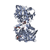
| Entry | Database: PDB / ID: 3nb0 | ||||||
|---|---|---|---|---|---|---|---|
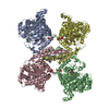
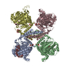
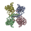
| Title | Glucose-6-Phosphate activated form of Yeast Glycogen Synthase | ||||||
 Components Components | Glycogen [starch] synthase isoform 2 | ||||||
 Keywords Keywords | TRANSFERASE / Glycogen Synthase / Glucose-6-phosphate / Yeast / Allosteric activation | ||||||
| Function / homology |  Function and homology information Function and homology informationGlycogen synthesis / glycogen binding / glycogen(starch) synthase / alpha-1,4-glucan glucosyltransferase (UDP-glucose donor) activity / glycogen granule / glycogen biosynthetic process / identical protein binding / nucleus / cytoplasm / cytosol Similarity search - Function | ||||||
| Biological species |  | ||||||
| Method |  X-RAY DIFFRACTION / SYNCHROTRON / MOLECULAR REPLACEMENT / Resolution: 2.406 Å X-RAY DIFFRACTION / SYNCHROTRON / MOLECULAR REPLACEMENT / Resolution: 2.406 Å | ||||||
 Authors Authors | Baskaran, S. / Hurley, T.D. | ||||||
 Citation Citation | Journal: Proc.Natl.Acad.Sci.USA / Year: 2010 Title: Structural basis for glucose-6-phosphate activation of glycogen synthase. Authors: Baskaran, S. / Roach, P.J. / Depaoli-Roach, A.A. / Hurley, T.D. | ||||||
| History |
|
- Structure visualization
| Structure viewer | Molecule: MolmilJmol/JSmol |
|---|
- Downloads & links
-Download
| PDBx/mmCIF format | 3nb0.cif.gz | 521.3 KB | Display | PDBx/mmCIF format |
|---|---|---|---|---|
| PDB format | pdb3nb0.ent.gz | 428.5 KB | Display | PDB format |
| PDBx/mmJSON format | 3nb0.json.gz | Tree view | PDBx/mmJSON format | |
| Others |  Other downloads Other downloads |
-Validation report
| Arichive directory | https://data.pdbj.org/pub/pdb/validation_reports/nb/3nb0ftp://data.pdbj.org/pub/pdb/validation_reports/nb/3nb0 | HTTPS FTP |
|---|
-Related structure data
| Related structure data |  3nazSC  3nchC  3o3cC S: Starting model for refinement C: citing same article ( |
|---|---|
| Similar structure data |


-Links
 PDBj
PDBj- Assembly
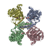
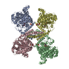
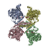
| Deposited unit | 
| ||||||||
|---|---|---|---|---|---|---|---|---|---|
| 1 |
| ||||||||
| Unit cell |
|
-Components
| #1: Protein | Mass: 82198.867 Da / Num. of mol.: 4 / Mutation: R589A R592A Source method: isolated from a genetically manipulated source Source: (gene. exp.) Gene: GSY2, L8479.8, YLR258W / Plasmid: pET28a / Production host:  #2: Sugar | ChemComp-G6P / 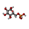  Type: D-saccharide, alpha linking / Mass: 260.136 Da / Num. of mol.: 6 Type: D-saccharide, alpha linking / Mass: 260.136 Da / Num. of mol.: 6Source method: isolated from a genetically manipulated source Formula: C6H13O9P #3: Chemical | ChemComp-PEG /   Mass: 106.120 Da / Num. of mol.: 6 / Source method: obtained synthetically / Formula: C4H10O3 Mass: 106.120 Da / Num. of mol.: 6 / Source method: obtained synthetically / Formula: C4H10O3#4: Water | ChemComp-HOH / |  Mass: 18.015 Da / Num. of mol.: 524 / Source method: isolated from a natural source / Formula: H2O Mass: 18.015 Da / Num. of mol.: 524 / Source method: isolated from a natural source / Formula: H2OSequence details | THE CRYSTALLIZED SEQUENCE HAS BEEN REFERENCED TO UNP ENTRY P27472. HOWEVER, RESIDUE A535S IS ...THE CRYSTALLIZ | |
|---|
-Experimental details
-Experiment
| Experiment | Method: X-RAY DIFFRACTION / Number of used crystals: 1 |
|---|
- Sample preparation
| Crystal | Density Matthews: 3.12 Å3/Da / Density % sol: 60.6 % |
|---|---|
| Crystal grow | Temperature: 293 K / Method: vapor diffusion, hanging drop / pH: 6.2 Details: 0.1M Bis-Tris pH 6.2, 25% PEG 300, VAPOR DIFFUSION, HANGING DROP, temperature 293K |
-Data collection
| Diffraction | Mean temperature: 100 K |
|---|---|
| Diffraction source | Source: SYNCHROTRON / Site: APS  / Beamline: 23-ID-D / Wavelength: 0.979 Å / Beamline: 23-ID-D / Wavelength: 0.979 Å |
| Detector | Type: MARMOSAIC 300 mm CCD / Detector: CCD / Date: Mar 14, 2010 |
| Radiation | Protocol: SINGLE WAVELENGTH / Monochromatic (M) / Laue (L): M / Scattering type: x-ray |
| Radiation wavelength | Wavelength: 0.979 Å / Relative weight: 1 |
| Reflection | Resolution: 2.4→50 Å / Num. all: 159580 / Num. obs: 157186 / % possible obs: 98.5 % / Observed criterion σ(F): 0.2 / Observed criterion σ(I): 0.2 / Redundancy: 4.8 % / Biso Wilson estimate: 42.44 Å2 / Rmerge(I) obs: 0.087 / Χ2: 1.062 / Net I/σ(I): 15.5 |
| Reflection shell | Resolution: 2.4→2.44 Å / Redundancy: 3.1 % / Rmerge(I) obs: 0.552 / Num. unique all: 6244 / Χ2: 1.092 / % possible all: 79.1 |
- Processing
| Software |
| |||||||||||||||||||||||||||||||||||||||||||||||||||||||||||||||||||||||||||||||||||||||||||||||||||||||||||||||||||||||||||||||||||||||||||||||||||||||||||||||||||||||||||||||||||||||||||||||||||||||||||||||||||||||||
|---|---|---|---|---|---|---|---|---|---|---|---|---|---|---|---|---|---|---|---|---|---|---|---|---|---|---|---|---|---|---|---|---|---|---|---|---|---|---|---|---|---|---|---|---|---|---|---|---|---|---|---|---|---|---|---|---|---|---|---|---|---|---|---|---|---|---|---|---|---|---|---|---|---|---|---|---|---|---|---|---|---|---|---|---|---|---|---|---|---|---|---|---|---|---|---|---|---|---|---|---|---|---|---|---|---|---|---|---|---|---|---|---|---|---|---|---|---|---|---|---|---|---|---|---|---|---|---|---|---|---|---|---|---|---|---|---|---|---|---|---|---|---|---|---|---|---|---|---|---|---|---|---|---|---|---|---|---|---|---|---|---|---|---|---|---|---|---|---|---|---|---|---|---|---|---|---|---|---|---|---|---|---|---|---|---|---|---|---|---|---|---|---|---|---|---|---|---|---|---|---|---|---|---|---|---|---|---|---|---|---|---|---|---|---|---|---|---|---|
| Refinement | Method to determine structure: MOLECULAR REPLACEMENT Starting model: PDB ENTRY 3NAZ Resolution: 2.406→46.229 Å / Occupancy max: 1 / Occupancy min: 0.4 / FOM work R set: 0.816 / SU ML: 2.37 / Isotropic thermal model: Isotropic / Cross valid method: Thoughout / σ(F): 0.06 / Phase error: 25.55 / Stereochemistry target values: ML
| |||||||||||||||||||||||||||||||||||||||||||||||||||||||||||||||||||||||||||||||||||||||||||||||||||||||||||||||||||||||||||||||||||||||||||||||||||||||||||||||||||||||||||||||||||||||||||||||||||||||||||||||||||||||||
| Solvent computation | Shrinkage radii: 0.9 Å / VDW probe radii: 1.11 Å / Solvent model: FLAT BULK SOLVENT MODEL / Bsol: 58.359 Å2 / ksol: 0.356 e/Å3 | |||||||||||||||||||||||||||||||||||||||||||||||||||||||||||||||||||||||||||||||||||||||||||||||||||||||||||||||||||||||||||||||||||||||||||||||||||||||||||||||||||||||||||||||||||||||||||||||||||||||||||||||||||||||||
| Displacement parameters | Biso max: 115.97 Å2 / Biso mean: 57.191 Å2 / Biso min: 23.14 Å2
| |||||||||||||||||||||||||||||||||||||||||||||||||||||||||||||||||||||||||||||||||||||||||||||||||||||||||||||||||||||||||||||||||||||||||||||||||||||||||||||||||||||||||||||||||||||||||||||||||||||||||||||||||||||||||
| Refinement step | Cycle: LAST / Resolution: 2.406→46.229 Å
| |||||||||||||||||||||||||||||||||||||||||||||||||||||||||||||||||||||||||||||||||||||||||||||||||||||||||||||||||||||||||||||||||||||||||||||||||||||||||||||||||||||||||||||||||||||||||||||||||||||||||||||||||||||||||
| Refine LS restraints |
| |||||||||||||||||||||||||||||||||||||||||||||||||||||||||||||||||||||||||||||||||||||||||||||||||||||||||||||||||||||||||||||||||||||||||||||||||||||||||||||||||||||||||||||||||||||||||||||||||||||||||||||||||||||||||
| LS refinement shell | Refine-ID: X-RAY DIFFRACTION / Total num. of bins used: 30
|
