 Movie
Movie Controller

[English] 日本語
 Yorodumi
- PDB-3n2t: Structure of the glycerol dehydrogenase AKR11B4 from Gluconobacte... -
+ Open data

- Basic information
| Entry | Database: PDB / ID: 3n2t | ||||||
|---|---|---|---|---|---|---|---|
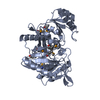
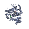
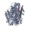
| Title | Structure of the glycerol dehydrogenase AKR11B4 from Gluconobacter oxydans | ||||||
 Components Components | Putative oxidoreductase | ||||||
 Keywords Keywords | OXIDOREDUCTASE / ALDO/KETO Reductase superfamily / AKR / AKR11B4 / TIM BARREL | ||||||
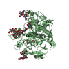
| Function / homology |  Function and homology information Function and homology informationOxidoreductases; Acting on the CH-OH group of donors; With NAD+ or NADP+ as acceptor / oxidoreductase activity / cytosol Similarity search - Function | ||||||
| Biological species |  Gluconobacter oxydans (bacteria) Gluconobacter oxydans (bacteria) | ||||||
| Method |  X-RAY DIFFRACTION / SYNCHROTRON / MOLECULAR REPLACEMENT / Resolution: 2 Å X-RAY DIFFRACTION / SYNCHROTRON / MOLECULAR REPLACEMENT / Resolution: 2 Å | ||||||
 Authors Authors | Richter, N. / Breicha, K. / Hummel, W. / Niefind, K. | ||||||
 Citation Citation | Journal: J.Mol.Biol. / Year: 2010 Title: The Three-Dimensional Structure of AKR11B4, a Glycerol Dehydrogenase from Gluconobacter oxydans, Reveals a Tryptophan Residue as an Accelerator of Reaction Turnover. Authors: Richter, N. / Breicha, K. / Hummel, W. / Niefind, K. #1: Journal: Chembiochem / Year: 2009 Title: Characterisation of a recombinant NADP-dependent glycerol dehydrogenase from Gluconobacter oxydans and its application in the production of L-glyceraldehyde. Authors: Richter, N. / Neumann, M. / Liese, A. / Wohlgemuth, R. / Eggert, T. / Hummel, W. | ||||||
| History |
|
- Structure visualization
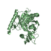
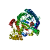
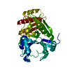
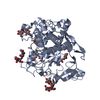
| Structure viewer | Molecule: MolmilJmol/JSmol |
|---|
- Downloads & links
-Download
| PDBx/mmCIF format | 3n2t.cif.gz | 80 KB | Display | PDBx/mmCIF format |
|---|---|---|---|---|
| PDB format | pdb3n2t.ent.gz | 59.1 KB | Display | PDB format |
| PDBx/mmJSON format | 3n2t.json.gz | Tree view | PDBx/mmJSON format | |
| Others |  Other downloads Other downloads |
-Validation report
| Arichive directory | https://data.pdbj.org/pub/pdb/validation_reports/n2/3n2tftp://data.pdbj.org/pub/pdb/validation_reports/n2/3n2t | HTTPS FTP |
|---|
-Related structure data
| Similar structure data |
|---|


-Links
 PDBj
PDBj
- Assembly
| Deposited unit | 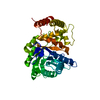
| ||||||||
|---|---|---|---|---|---|---|---|---|---|
| 1 |
| ||||||||
| Unit cell |
|
-Components
| #1: Protein | Mass: 39076.410 Da / Num. of mol.: 1 Source method: isolated from a genetically manipulated source Source: (gene. exp.) Gluconobacter oxydans (bacteria) / Gene: GOX1615 / Plasmid: pET28a (NdeI/SalI) / Production host: References: UniProt: Q5FQJ0, Oxidoreductases; Acting on the CH-OH group of donors; With NAD+ or NADP+ as acceptor |
|---|---|
| #2: Water | ChemComp-HOH /  Mass: 18.015 Da / Num. of mol.: 150 / Source method: isolated from a natural source / Formula: H2O Mass: 18.015 Da / Num. of mol.: 150 / Source method: isolated from a natural source / Formula: H2O |
-Experimental details
-Experiment
| Experiment | Method: X-RAY DIFFRACTION |
|---|
- Sample preparation
| Crystal | Density Matthews: 1.97 Å3/Da / Density % sol: 37.56 % |
|---|---|
| Crystal grow | Temperature: 293 K / Method: vapor diffusion, sitting drop Details: protein stock solution: 10 mg/ml AKR11B4 protein, 10 mM Tris/HCl puffer, 150 mM NaCl, 0.5 mM EDTA, pH 8.5. Reservoir solution: 35 % (w/v) poly ethylen glycol 3350 (PEG 3350), 200 mM ...Details: protein stock solution: 10 mg/ml AKR11B4 protein, 10 mM Tris/HCl puffer, 150 mM NaCl, 0.5 mM EDTA, pH 8.5. Reservoir solution: 35 % (w/v) poly ethylen glycol 3350 (PEG 3350), 200 mM potassium nitrate. The crystallization drop contained equal volumes of the reservoir and the protein stock solution before equilibration. The pH-value in the crystallization drop was determined by the buffer of the protein stock solution., VAPOR DIFFUSION, SITTING DROP, temperature 293K |
-Data collection
| Diffraction source | Source: SYNCHROTRON / Site: EMBL/DESY, HAMBURG  / Beamline: X11 / Wavelength: 0.9537 Å / Beamline: X11 / Wavelength: 0.9537 Å |
|---|---|
| Radiation | Protocol: SINGLE WAVELENGTH / Monochromatic (M) / Laue (L): M / Scattering type: x-ray |
| Radiation wavelength | Wavelength: 0.9537 Å / Relative weight: 1 |
| Reflection | Resolution: 2→34.5 Å / Num. obs: 26999 / % possible obs: 99.8 % / Redundancy: 5.4 % / Biso Wilson estimate: 29.1 Å2 / Rmerge(I) obs: 0.133 / Rsym value: 0.133 / Net I/σ(I): 10.1 |
- Processing
| Software |
| ||||||||||||||||||||||||||||||||||||||||||||||||||||||||
|---|---|---|---|---|---|---|---|---|---|---|---|---|---|---|---|---|---|---|---|---|---|---|---|---|---|---|---|---|---|---|---|---|---|---|---|---|---|---|---|---|---|---|---|---|---|---|---|---|---|---|---|---|---|---|---|---|---|
| Refinement | Method to determine structure: MOLECULAR REPLACEMENT / Resolution: 2→24.119 Å / SU ML: 0.24 / σ(F): 1.37 / Stereochemistry target values: ML
| ||||||||||||||||||||||||||||||||||||||||||||||||||||||||
| Solvent computation | Shrinkage radii: 0.9 Å / VDW probe radii: 1.11 Å / Solvent model: FLAT BULK SOLVENT MODEL / Bsol: 28.413 Å2 / ksol: 0.344 e/Å3 | ||||||||||||||||||||||||||||||||||||||||||||||||||||||||
| Displacement parameters |
| ||||||||||||||||||||||||||||||||||||||||||||||||||||||||
| Refinement step | Cycle: LAST / Resolution: 2→24.119 Å
| ||||||||||||||||||||||||||||||||||||||||||||||||||||||||
| Refine LS restraints |
| ||||||||||||||||||||||||||||||||||||||||||||||||||||||||
| LS refinement shell |
|
