 Movie
Movie Controller

[English] 日本語
 Yorodumi
- PDB-3mwu: Activated Calcium-Dependent Protein Kinase 1 from Cryptosporidium... -
+ Open data

- Basic information
| Entry | Database: PDB / ID: 3mwu | ||||||
|---|---|---|---|---|---|---|---|
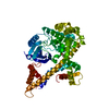
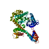
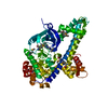
| Title | Activated Calcium-Dependent Protein Kinase 1 from Cryptosporidium parvum (CpCDPK1) in complex with bumped kinase inhibitor RM-1-95 | ||||||
 Components Components | Calmodulin-domain protein kinase 1 | ||||||
 Keywords Keywords | TRANSFERASE/TRANSFERASE INHIBITOR / serine/threonine protein kinase / transferase / calcium-binding / ATP-binding / calmodulin / bumped kinase inhibitor / BKI / Structural Genomics / Medical Structural Genomics of Pathogenic Protozoa / MSGPP / TRANSFERASE-TRANSFERASE INHIBITOR complex | ||||||
| Function / homology |  Function and homology information Function and homology informationnon-specific serine/threonine protein kinase / protein serine/threonine kinase activity / calcium ion binding / ATP binding Similarity search - Function | ||||||
| Biological species |   Cryptosporidium parvum (eukaryote) Cryptosporidium parvum (eukaryote) | ||||||
| Method |  X-RAY DIFFRACTION / SYNCHROTRON / FOURIER SYNTHESIS / Resolution: 1.98 Å X-RAY DIFFRACTION / SYNCHROTRON / FOURIER SYNTHESIS / Resolution: 1.98 Å | ||||||
 Authors Authors | Larson, E.T. / Merritt, E.A. / Medical Structural Genomics of Pathogenic Protozoa (MSGPP) | ||||||
 Citation Citation | Journal: ACS Med Chem Lett / Year: 2010 Title: Discovery of Potent and Selective Inhibitors of Calcium-Dependent Protein Kinase 1 (CDPK1) from C. parvum and T. gondii. Authors: Murphy, R.C. / Ojo, K.K. / Larson, E.T. / Castellanos-Gonzalez, A. / Perera, B.G. / Keyloun, K.R. / Kim, J.E. / Bhandari, J.G. / Muller, N.R. / Verlinde, C.L. / White, A.C. / Merritt, E.A. / ...Authors: Murphy, R.C. / Ojo, K.K. / Larson, E.T. / Castellanos-Gonzalez, A. / Perera, B.G. / Keyloun, K.R. / Kim, J.E. / Bhandari, J.G. / Muller, N.R. / Verlinde, C.L. / White, A.C. / Merritt, E.A. / Van Voorhis, W.C. / Maly, D.J. #1: Journal: Nat.Struct.Mol.Biol. / Year: 2010Title: Toxoplasma gondii calcium-dependent protein kinase 1 is a target for selective kinase inhibitors. Authors: Ojo, K.K. / Larson, E.T. / Keyloun, K.R. / Castaneda, L.J. / Derocher, A.E. / Inampudi, K.K. / Kim, J.E. / Arakaki, T.L. / Murphy, R.C. / Zhang, L. / Napuli, A.J. / Maly, D.J. / Verlinde, C. ...Authors: Ojo, K.K. / Larson, E.T. / Keyloun, K.R. / Castaneda, L.J. / Derocher, A.E. / Inampudi, K.K. / Kim, J.E. / Arakaki, T.L. / Murphy, R.C. / Zhang, L. / Napuli, A.J. / Maly, D.J. / Verlinde, C.L. / Buckner, F.S. / Parsons, M. / Hol, W.G. / Merritt, E.A. / Van Voorhis, W.C. | ||||||
| History |
|
- Structure visualization
| Structure viewer | Molecule: MolmilJmol/JSmol |
|---|
- Downloads & links
-Download
| PDBx/mmCIF format | 3mwu.cif.gz | 204.9 KB | Display | PDBx/mmCIF format |
|---|---|---|---|---|
| PDB format | pdb3mwu.ent.gz | 162.5 KB | Display | PDB format |
| PDBx/mmJSON format | 3mwu.json.gz | Tree view | PDBx/mmJSON format | |
| Others |  Other downloads Other downloads |
-Validation report
| Arichive directory | https://data.pdbj.org/pub/pdb/validation_reports/mw/3mwuftp://data.pdbj.org/pub/pdb/validation_reports/mw/3mwu | HTTPS FTP |
|---|
-Related structure data
| Related structure data |  3n51C  3ncgC 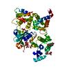 3igoS C: citing same article ( S: Starting model for refinement |
|---|---|
| Similar structure data |

-Links
 PDBj
PDBj
- Assembly
| Deposited unit | 
| ||||||||
|---|---|---|---|---|---|---|---|---|---|
| 1 |
| ||||||||
| Unit cell |
|
-Components
| #1: Protein | Mass: 56152.793 Da / Num. of mol.: 1 / Fragment: CpCDPK1 Mutation: N-terminal 69 residues replaced with His tag and linker Source method: isolated from a genetically manipulated source Source: (gene. exp.) Cryptosporidium parvum (eukaryote) / Strain: Iowa II / Gene: CDPK1, cgd3_920 / Plasmid: PET15MHL / Production host:  References: UniProt: A3FQ16, Ca2+/calmodulin-dependent protein kinase | ||||||
|---|---|---|---|---|---|---|---|
| #2: Chemical | ChemComp-CA /   Mass: 40.078 Da / Num. of mol.: 4 / Source method: obtained synthetically / Formula: Ca Mass: 40.078 Da / Num. of mol.: 4 / Source method: obtained synthetically / Formula: Ca#3: Chemical | ChemComp-BK3 / |   Mass: 372.466 Da / Num. of mol.: 1 / Source method: obtained synthetically / Formula: C22H24N6 Mass: 372.466 Da / Num. of mol.: 1 / Source method: obtained synthetically / Formula: C22H24N6#4: Water | ChemComp-HOH / |  Mass: 18.015 Da / Num. of mol.: 125 / Source method: isolated from a natural source / Formula: H2O Mass: 18.015 Da / Num. of mol.: 125 / Source method: isolated from a natural source / Formula: H2OHas protein modification | N | |
-Experimental details
-Experiment
| Experiment | Method: X-RAY DIFFRACTION / Number of used crystals: 1 |
|---|
- Sample preparation
| Crystal | Density Matthews: 2.38 Å3/Da / Density % sol: 48.27 % |
|---|---|
| Crystal grow | Temperature: 298 K / pH: 7 Details: 27% PEG 3350, 0.27 M ammonium tartrate (pH 7.0), 6% PEG 400, 5 mM TCEP, 4 mM MgCl2, 2 mM CaCl2, 2 mM inhibitor, VAPOR DIFFUSION, SITTING DROP, temperature 298K |
-Data collection
| Diffraction | Mean temperature: 100 K |
|---|---|
| Diffraction source | Source: SYNCHROTRON / Site: SSRL  / Beamline: BL9-2 / Wavelength: 0.9797 / Beamline: BL9-2 / Wavelength: 0.9797 |
| Detector | Type: MARMOSAIC 325 mm CCD / Detector: CCD / Date: Feb 4, 2010 |
| Radiation | Monochromator: DOUBLE CRYSTAL / Protocol: SINGLE WAVELENGTH / Monochromatic (M) / Laue (L): M / Scattering type: x-ray |
| Radiation wavelength | Wavelength: 0.9797 Å / Relative weight: 1 |
| Reflection | Resolution: 1.98→58.03 Å / Num. obs: 36138 / % possible obs: 99.8 % / Observed criterion σ(I): 5 / Redundancy: 3.71 % / Biso Wilson estimate: 34.8 Å2 / Rmerge(I) obs: 0.046 / Net I/σ(I): 11.1548 |
| Reflection shell | Resolution: 1.98→2.1 Å / Redundancy: 3.67 % / Rmerge(I) obs: 0.43 / Mean I/σ(I) obs: 2.3 / % possible all: 99.8 |
- Processing
| Software |
| ||||||||||||||||||||||||||||||||||||||||||||||||||||||||||||||||||||||||||||||||||||||||||||||||||||||||||||||||||||||||||||||||||||||||||||||||||||||||||||||||||||||||||
|---|---|---|---|---|---|---|---|---|---|---|---|---|---|---|---|---|---|---|---|---|---|---|---|---|---|---|---|---|---|---|---|---|---|---|---|---|---|---|---|---|---|---|---|---|---|---|---|---|---|---|---|---|---|---|---|---|---|---|---|---|---|---|---|---|---|---|---|---|---|---|---|---|---|---|---|---|---|---|---|---|---|---|---|---|---|---|---|---|---|---|---|---|---|---|---|---|---|---|---|---|---|---|---|---|---|---|---|---|---|---|---|---|---|---|---|---|---|---|---|---|---|---|---|---|---|---|---|---|---|---|---|---|---|---|---|---|---|---|---|---|---|---|---|---|---|---|---|---|---|---|---|---|---|---|---|---|---|---|---|---|---|---|---|---|---|---|---|---|---|---|---|
| Refinement | Method to determine structure: FOURIER SYNTHESIS Starting model: 3IGO, PROTEIN ONLY Resolution: 1.98→42.04 Å / Cor.coef. Fo:Fc: 0.957 / Cor.coef. Fo:Fc free: 0.942 / Occupancy max: 1 / Occupancy min: 0.45 / SU B: 8.394 / SU ML: 0.122 / Cross valid method: THROUGHOUT / σ(F): 0 / ESU R: 0.175 / ESU R Free: 0.154 / Stereochemistry target values: MAXIMUM LIKELIHOOD Details: HYDROGENS HAVE BEEN ADDED IN THE RIDING POSITIONS U VALUES : WITH TLS ADDED
| ||||||||||||||||||||||||||||||||||||||||||||||||||||||||||||||||||||||||||||||||||||||||||||||||||||||||||||||||||||||||||||||||||||||||||||||||||||||||||||||||||||||||||
| Solvent computation | Ion probe radii: 0.8 Å / Shrinkage radii: 0.8 Å / VDW probe radii: 1.4 Å / Solvent model: BABINET MODEL WITH MASK | ||||||||||||||||||||||||||||||||||||||||||||||||||||||||||||||||||||||||||||||||||||||||||||||||||||||||||||||||||||||||||||||||||||||||||||||||||||||||||||||||||||||||||
| Displacement parameters | Biso mean: 55.41 Å2
| ||||||||||||||||||||||||||||||||||||||||||||||||||||||||||||||||||||||||||||||||||||||||||||||||||||||||||||||||||||||||||||||||||||||||||||||||||||||||||||||||||||||||||
| Refinement step | Cycle: LAST / Resolution: 1.98→42.04 Å
| ||||||||||||||||||||||||||||||||||||||||||||||||||||||||||||||||||||||||||||||||||||||||||||||||||||||||||||||||||||||||||||||||||||||||||||||||||||||||||||||||||||||||||
| Refine LS restraints |
| ||||||||||||||||||||||||||||||||||||||||||||||||||||||||||||||||||||||||||||||||||||||||||||||||||||||||||||||||||||||||||||||||||||||||||||||||||||||||||||||||||||||||||
| LS refinement shell | Resolution: 1.98→2.03 Å / Total num. of bins used: 20
| ||||||||||||||||||||||||||||||||||||||||||||||||||||||||||||||||||||||||||||||||||||||||||||||||||||||||||||||||||||||||||||||||||||||||||||||||||||||||||||||||||||||||||
| Refinement TLS params. | Method: refined / Refine-ID: X-RAY DIFFRACTION
| ||||||||||||||||||||||||||||||||||||||||||||||||||||||||||||||||||||||||||||||||||||||||||||||||||||||||||||||||||||||||||||||||||||||||||||||||||||||||||||||||||||||||||
| Refinement TLS group |
|
