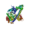
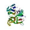
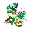
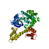
- PDB-3i79: Calcium-Dependent Protein Kinase 1 from Toxoplasma gondii (TgCDPK1) -
+
Open data
ID or keywords:
Loading...
-
Basic information
Entry
Database: PDB / ID: 3i79
Title
Calcium-Dependent Protein Kinase 1 from Toxoplasma gondii (TgCDPK1)
Components
Calmodulin-domain protein kinase 1
Keywords
TRANSFERASE / protein kinase / calmodulin / EF hand / bumped kinase inhibitor / ATP-binding / Kinase / Nucleotide-binding / Serine/threonine-protein kinase / Structural Genomics / Medical Structural Genomics of Pathogenic Protozoa / MSGPP
Function / homology
Function and homology information
non-specific serine/threonine protein kinase / protein serine/threonine kinase activity / calcium ion binding / ATP binding Similarity search - Function
Mass: 55226.914 Da / Num. of mol.: 1 / Fragment: residues 30-507 Source method: isolated from a genetically manipulated source Details: residues 1-29 were replaced with a 3C cleavable His-tag and linker during cloning; tag was cleaved prior to crystallization Source: (gene. exp.) Toxoplasma gondii (eukaryote) / Gene: CDPK1 / Plasmid: AVA0421 / Production host: Escherichia coli (E. coli) / Strain (production host): BL21(DE3) References: UniProt: Q9BJF5, Ca2+/calmodulin-dependent protein kinase
Mass: 18.015 Da / Num. of mol.: 75 / Source method: isolated from a natural source / Formula: H2O
-
Experimental details
-
Experiment
Experiment
Method: X-RAY DIFFRACTION / Number of used crystals: 1
-
Sample preparation
Crystal
Density Matthews: 2.05 Å3/Da / Density % sol: 40.09 %
Crystal grow
Temperature: 298 K / Method: sitting drop vapor diffusion / pH: 6.5 Details: 25% PEG 3350, 0.2 M tri-ammonium citrate; cryoprotected with 15% ethylene glycol, pH 6.5, sitting drop vapor diffusion, temperature 298K
-
Data collection
Diffraction
ID
Mean temperature (K)
Crystal-ID
1
100
1
2
100
1
1,2
1
Diffraction source
Source
Site
Beamline
ID
Wavelength (Å)
SYNCHROTRON
SSRL
BL9-2
1
0.97923
SYNCHROTRON
SSRL
BL9-2
2
0.97923
Detector
Type
ID
Detector
Date
MarUSA MarMosaic -325 CCD
1
CCD
Apr 22, 2009
MarUSA MarMosaic -325 CCD
2
CCD
Apr 22, 2009
Radiation
ID
Monochromator
Protocol
Scattering type
Wavelength-ID
1
Doublecrystal
SAD
x-ray
1
2
Doublecrystal
SINGLEWAVELENGTH
x-ray
1
Radiation wavelength
Wavelength: 0.97923 Å / Relative weight: 1
Reflection
Redundancy: 7.7 % / Av σ(I) over netI: 27.54 / Number: 154144 / Rmerge(I) obs: 0.109 / Χ2: 1.03 / D res high: 2.3 Å / D res low: 50 Å / Num. obs: 20099 / % possible obs: 100
Diffraction reflection shell
Highest resolution (Å)
Lowest resolution (Å)
% possible obs (%)
ID
Rmerge(I) obs
Chi squared
Redundancy
4.95
50
99.8
1
0.05
1.009
7.5
3.93
4.95
100
1
0.049
1.107
7.7
3.44
3.93
100
1
0.074
1.088
7.7
3.12
3.44
100
1
0.113
1.082
7.7
2.9
3.12
100
1
0.183
1.06
7.7
2.73
2.9
100
1
0.3
1.04
7.7
2.59
2.73
100
1
0.451
0.987
7.7
2.48
2.59
100
1
0.647
0.982
7.7
2.38
2.48
100
1
0.785
0.971
7.7
2.3
2.38
100
1
0.954
0.969
7.7
Reflection
Resolution: 2.04→50 Å / Num. obs: 28324 / % possible obs: 99.9 % / Observed criterion σ(I): 3 / Redundancy: 4.2 % / Biso Wilson estimate: 34.8 Å2 / Rmerge(I) obs: 0.052 / Χ2: 1.04 / Net I/σ(I): 10.4
Reflection shell
Resolution: 2.04→2.12 Å / Redundancy: 4.2 % / Rmerge(I) obs: 0.67 / Mean I/σ(I) obs: 2.2 / Num. unique all: 2799 / Χ2: 1.041 / % possible all: 100
-
Phasing
Phasing
Method: SAD
Phasing MAD
D res high: 2.68 Å / D res low: 50 Å / FOM : 0.34 / Reflection: 12526
In the structure databanks used in Yorodumi, some data are registered as the other names, "COVID-19 virus" and "2019-nCoV". Here are the details of the virus and the list of structure data.
Jan 31, 2019. EMDB accession codes are about to change! (news from PDBe EMDB page)
EMDB accession codes are about to change! (news from PDBe EMDB page)
The allocation of 4 digits for EMDB accession codes will soon come to an end. Whilst these codes will remain in use, new EMDB accession codes will include an additional digit and will expand incrementally as the available range of codes is exhausted. The current 4-digit format prefixed with “EMD-” (i.e. EMD-XXXX) will advance to a 5-digit format (i.e. EMD-XXXXX), and so on. It is currently estimated that the 4-digit codes will be depleted around Spring 2019, at which point the 5-digit format will come into force.
The EM Navigator/Yorodumi systems omit the EMD- prefix.
Related info.:Q: What is EMD? / ID/Accession-code notation in Yorodumi/EM Navigator
Yorodumi is a browser for structure data from EMDB, PDB, SASBDB, etc.
This page is also the successor to EM Navigator detail page, and also detail information page/front-end page for Omokage search.
The word "yorodu" (or yorozu) is an old Japanese word meaning "ten thousand". "mi" (miru) is to see.
Related info.:EMDB / PDB / SASBDB / Comparison of 3 databanks / Yorodumi Search / Aug 31, 2016. New EM Navigator & Yorodumi / Yorodumi Papers / Jmol/JSmol / Function and homology information / Changes in new EM Navigator and Yorodumi
 Movie
Movie Controller

 Yorodumi
 Open data
 Basic information
 Components
 Keywords
 Function and homology information
 X-RAY DIFFRACTION /
  Authors
 Citation
 Structure visualization
 Downloads & links
 Other downloads
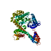


 PDBj
PDBj
 Assembly


 Mass: 62.068 Da / Num. of mol.: 1 / Source method: obtained synthetically / Formula: C2H6O2
 Mass: 18.015 Da / Num. of mol.: 75 / Source method: isolated from a natural source / Formula: H2O
 Sample preparation
 Processing