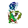
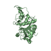
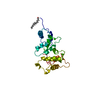
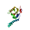
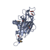
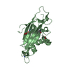
Peptidase M73, camelysin / Camelysin metallo-endopeptidase / Signal peptide, camelysin / sporulation resulting in formation of a cellular spore / extracellular region / identical protein binding / 2-HYDROXYBENZOIC ACID / Major biofilm matrix component
Function and homology information
Biological species
Bacillus subtilis (bacteria)
Method
X-RAY DIFFRACTION / SYNCHROTRON / SAD / Resolution: 1.56 Å
Protocol: SINGLE WAVELENGTH / Monochromatic (M) / Laue (L): M / Scattering type: x-ray
Radiation wavelength
Wavelength: 0.9797 Å / Relative weight: 1
Reflection twin
Crystal-ID
ID
Operator
Domain-ID
Fraction
1
1
H, K, L
1
0.524
1
1
-H, K, -L
2
0.476
Reflection
Resolution: 1.56→40.71 Å / Num. obs: 47368 / % possible obs: 96.33 % / Redundancy: 4.1 % / Biso Wilson estimate: 24.363 Å2 / CC1/2: 0.996 / Rmerge(I) obs: 0.0777 / Net I/σ(I): 13.33
Reflection shell
Resolution: 1.56→1.614 Å / Redundancy: 4.2 % / Rmerge(I) obs: 0.4156 / Mean I/σ(I) obs: 2.73 / Num. unique obs: 4658 / CC1/2: 0.921 / % possible all: 93.31
-
Processing
Software
Name
Version
Classification
REFMAC
5.8.0151
refinement
XDS
datareduction
XDS
datascaling
AutoSol
phasing
Refinement
Method to determine structure: SAD / Resolution: 1.56→40.71 Å / Cor.coef. Fo:Fc: 0.973 / Cor.coef. Fo:Fc free: 0.969 / SU B: 0.964 / SU ML: 0.037 / Cross valid method: THROUGHOUT / ESU R: 0.016 / ESU R Free: 0.016 / Details: HYDROGENS HAVE BEEN ADDED IN THE RIDING POSITIONS
Rfactor
Num. reflection
% reflection
Selection details
Rfree
0.17523
1080
2.2 %
RANDOM
Rwork
0.15222
-
-
-
obs
0.15273
47368
96.02 %
-
Solvent computation
Ion probe radii: 0.8 Å / Shrinkage radii: 0.8 Å / VDW probe radii: 1.2 Å
 Movie
Movie Controller

 Yorodumi
 Open data
 Basic information
 Components
 Keywords
 Function and homology information
 X-RAY DIFFRACTION /
  Authors
 Citation
 Structure visualization
 Downloads & links
 Other downloads
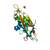


 PDBj
PDBj

 Assembly
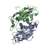
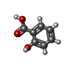
 Mass: 138.121 Da / Num. of mol.: 2 / Source method: obtained synthetically / Formula: C7H6O3
 Mass: 62.068 Da / Num. of mol.: 3 / Source method: obtained synthetically / Formula: C2H6O2
 Mass: 18.015 Da / Num. of mol.: 331 / Source method: isolated from a natural source / Formula: H2O
 Sample preparation
 / Beamline: 14.1 / Wavelength: 0.9797 Å
 Processing