- PDB-3mdo: Crystal structure of a Putative phosphoribosylformylglycinamidine... -
+
Open data
ID or keywords:
Loading...
-
Basic information
Entry
Database: PDB / ID: 3mdo
Title
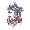
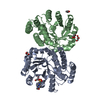
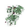
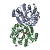
Crystal structure of a Putative phosphoribosylformylglycinamidine cyclo-ligase (BDI_2101) from Parabacteroides distasonis ATCC 8503 at 1.91 A resolution
Mass: 18.015 Da / Num. of mol.: 459 / Source method: isolated from a natural source / Formula: H2O
-
Details
Has protein modification
Y
Sequence details
THIS CONSTRUCT WAS EXPRESSED WITH A PURIFICATION TAG MGSDKIHHHHHHENLYFQG. THE TAG WAS REMOVED WITH ...THIS CONSTRUCT WAS EXPRESSED WITH A PURIFICATION TAG MGSDKIHHHHHHENLYFQG. THE TAG WAS REMOVED WITH TEV PROTEASE LEAVING ONLY A GLYCINE (0) FOLLOWED BY THE TARGET SEQUENCE.
-
Experimental details
-
Experiment
Experiment
Method: X-RAY DIFFRACTION / Number of used crystals: 1
-
Sample preparation
Crystal
Density Matthews: 2.36 Å3/Da / Density % sol: 47.78 % Description: DATA WERE SCALED USING XSCALE WITH FRIEDEL PAIRS KEPT AS SEPARATE WHEN COMPUTING R-SYM, COMPLETENESS AND
Resolution: 1.91→29.623 Å / Num. obs: 64592 / % possible obs: 99.7 % / Observed criterion σ(I): -3 / Biso Wilson estimate: 25.989 Å2 / Rmerge(I) obs: 0.074 / Net I/σ(I): 13.15
Reflection shell
Resolution (Å)
Rmerge(I) obs
Mean I/σ(I) obs
Num. measured obs
Num. unique obs
Diffraction-ID
% possible all
1.91-1.98
0.71
2
47459
12418
1
99.7
1.98-2.06
0.521
2.7
46945
12259
1
99.7
2.06-2.15
0.376
3.7
45020
11732
1
99.6
2.15-2.26
0.276
4.9
45737
11879
1
99.7
2.26-2.41
0.206
6.5
49826
12942
1
99.8
2.41-2.59
0.154
8.4
45718
11816
1
99.7
2.59-2.85
0.104
12.1
47263
12208
1
99.8
2.85-3.26
0.066
18.3
47421
12218
1
99.9
3.26-4.1
0.034
32
47544
12226
1
99.8
4.1-29.623
0.027
40.5
47908
12381
1
99.6
-
Phasing
Phasing
Method: MAD
-
Processing
Software
Name
Version
Classification
NB
REFMAC
5.5.0053
refinement
PHENIX
refinement
SHELX
phasing
MolProbity
3beta29
modelbuilding
XSCALE
datascaling
PDB_EXTRACT
3.006
dataextraction
XDS
datareduction
SHELXD
phasing
autoSHARP
phasing
Refinement
Method to determine structure: MAD / Resolution: 1.91→29.623 Å / Cor.coef. Fo:Fc: 0.97 / Cor.coef. Fo:Fc free: 0.956 / Occupancy max: 1 / Occupancy min: 0.25 / SU B: 5.816 / SU ML: 0.077 / TLS residual ADP flag: LIKELY RESIDUAL / Cross valid method: THROUGHOUT / σ(F): 0 / ESU R: 0.126 / ESU R Free: 0.119 Stereochemistry target values: MAXIMUM LIKELIHOOD WITH PHASES Details: 1. HYDROGENS HAVE BEEN ADDED IN THE RIDING POSITIONS. 2. A MET-INHIBITION PROTOCOL WAS USED FOR SELENOMETHIONINE INCORPORATION DURING PROTEIN EXPRESSION. THE OCCUPANCY OF THE SE ATOMS IN THE ...Details: 1. HYDROGENS HAVE BEEN ADDED IN THE RIDING POSITIONS. 2. A MET-INHIBITION PROTOCOL WAS USED FOR SELENOMETHIONINE INCORPORATION DURING PROTEIN EXPRESSION. THE OCCUPANCY OF THE SE ATOMS IN THE MSE RESIDUES WAS REDUCED TO 0.75 FOR THE REDUCED SCATTERING POWER DUE TO PARTIAL S-MET INCORPORATION. 3. ATOM RECORDS CONTAIN RESIDUAL B FACTORS ONLY. 4. REFMAC 5.5.0053 AUTOMATICALLY ASSIGNED WATERS TO EXISTING TLS GROUPS DURING REFINEMENT. 5. CALCIUM (CA), ACETATE (ACT), CACODYLATE (CAC) AND PEG FRAGMENTS (PEG, PGE, PG4 AND P6G) FROM THE CRYSTALLIZATION HAVE BEEN MODELED INTO THE STRUCTURE.
Rfactor
Num. reflection
% reflection
Selection details
Rfree
0.19
3265
5.1 %
RANDOM
Rwork
0.154
-
-
-
obs
0.156
64507
99.83 %
-
Solvent computation
Ion probe radii: 0.8 Å / Shrinkage radii: 0.8 Å / VDW probe radii: 1.4 Å / Solvent model: BABINET MODEL WITH MASK
In the structure databanks used in Yorodumi, some data are registered as the other names, "COVID-19 virus" and "2019-nCoV". Here are the details of the virus and the list of structure data.
Jan 31, 2019. EMDB accession codes are about to change! (news from PDBe EMDB page)
EMDB accession codes are about to change! (news from PDBe EMDB page)
The allocation of 4 digits for EMDB accession codes will soon come to an end. Whilst these codes will remain in use, new EMDB accession codes will include an additional digit and will expand incrementally as the available range of codes is exhausted. The current 4-digit format prefixed with “EMD-” (i.e. EMD-XXXX) will advance to a 5-digit format (i.e. EMD-XXXXX), and so on. It is currently estimated that the 4-digit codes will be depleted around Spring 2019, at which point the 5-digit format will come into force.
The EM Navigator/Yorodumi systems omit the EMD- prefix.
Related info.:Q: What is EMD? / ID/Accession-code notation in Yorodumi/EM Navigator
Yorodumi is a browser for structure data from EMDB, PDB, SASBDB, etc.
This page is also the successor to EM Navigator detail page, and also detail information page/front-end page for Omokage search.
The word "yorodu" (or yorozu) is an old Japanese word meaning "ten thousand". "mi" (miru) is to see.
Related info.:EMDB / PDB / SASBDB / Comparison of 3 databanks / Yorodumi Search / Aug 31, 2016. New EM Navigator & Yorodumi / Yorodumi Papers / Jmol/JSmol / Function and homology information / Changes in new EM Navigator and Yorodumi
 Movie
Movie Controller

 Yorodumi
 Open data
 Basic information
 Components
 Keywords
 Function and homology information
 Parabacteroides distasonis (bacteria)
 X-RAY DIFFRACTION /
  Authors
 Citation
 Structure visualization
 Downloads & links
 Other downloads


 PDBj
PDBj


 Assembly


 Mass: 40.078 Da / Num. of mol.: 7 / Source method: obtained synthetically / Formula: Ca
 Mass: 35.453 Da / Num. of mol.: 4 / Source method: obtained synthetically / Formula: Cl
 Mass: 59.044 Da / Num. of mol.: 2 / Source method: obtained synthetically / Formula: C2H3O2
 Mass: 106.120 Da / Num. of mol.: 11 / Source method: obtained synthetically / Formula: C4H10O3
 Mass: 150.173 Da / Num. of mol.: 5 / Source method: obtained synthetically / Formula: C6H14O4
 Mass: 282.331 Da / Num. of mol.: 1 / Source method: obtained synthetically / Formula: C12H26O7 / Comment: precipitant*YM
 Mass: 136.989 Da / Num. of mol.: 1 / Source method: obtained synthetically / Formula: C2H6AsO2
 Mass: 194.226 Da / Num. of mol.: 2 / Source method: obtained synthetically / Formula: C8H18O5 / Comment: precipitant*YM
 Sample preparation
 / Beamline: BL9-2 / Wavelength: 0.91837,0.97949
 Processing