 Movie
Movie Controller

[English] 日本語
 Yorodumi
- PDB-1s64: Rat protein geranylgeranyltransferase type-I complexed with L-778... -
+ Open data

- Basic information
| Entry | Database: PDB / ID: 1s64 | ||||||
|---|---|---|---|---|---|---|---|
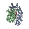
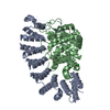
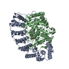
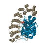
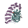
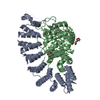
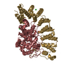
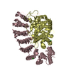
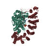
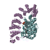
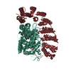
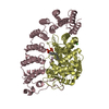
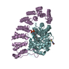
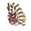
| Title | Rat protein geranylgeranyltransferase type-I complexed with L-778,123 and a sulfate anion | ||||||
 Components Components |
| ||||||
 Keywords Keywords | TRANSFERASE / L-778 / 123 / protein geranylgeranyltransferase type-I / protein prenylation / lipid modification / drug | ||||||
| Function / homology |  Function and homology information Function and homology informationApoptotic cleavage of cellular proteins / Inactivation, recovery and regulation of the phototransduction cascade / RAS processing / protein geranylgeranyltransferase activity / peptide pheromone maturation / protein geranylgeranyltransferase type I / CAAX-protein geranylgeranyltransferase activity / CAAX-protein geranylgeranyltransferase complex / protein farnesylation / protein farnesyltransferase ...Apoptotic cleavage of cellular proteins / Inactivation, recovery and regulation of the phototransduction cascade / RAS processing / protein geranylgeranyltransferase activity / peptide pheromone maturation / protein geranylgeranyltransferase type I / CAAX-protein geranylgeranyltransferase activity / CAAX-protein geranylgeranyltransferase complex / protein farnesylation / protein farnesyltransferase / protein farnesyltransferase activity / protein farnesyltransferase complex / Rab geranylgeranyltransferase activity / protein geranylgeranylation / nuclear envelope organization / acetyltransferase activator activity / positive regulation of skeletal muscle acetylcholine-gated channel clustering / heterocyclic compound binding / microtubule associated complex / enzyme-linked receptor protein signaling pathway / regulation of microtubule-based movement / alpha-tubulin binding / positive regulation of Rac protein signal transduction / positive regulation of cell cycle / protein maturation / receptor tyrosine kinase binding / microtubule binding / molecular adaptor activity / positive regulation of cell population proliferation / negative regulation of apoptotic process / enzyme binding / zinc ion binding / cytoplasm Similarity search - Function | ||||||
| Biological species |  | ||||||
| Method |  X-RAY DIFFRACTION / SYNCHROTRON / MOLECULAR REPLACEMENT / Resolution: 2.55 Å X-RAY DIFFRACTION / SYNCHROTRON / MOLECULAR REPLACEMENT / Resolution: 2.55 Å | ||||||
 Authors Authors | Reid, T.S. / Long, S.B. / Beese, L.S. | ||||||
 Citation Citation | Journal: Biochemistry / Year: 2004 Title: Crystallographic Analysis Reveals that Anticancer Clinical Candidate L-778,123 Inhibits Protein Farnesyltransferase and Geranylgeranyltransferase-I by Different Binding Modes. Authors: Reid, T.S. / Long, S.B. / Beese, L.S. | ||||||
| History |
|
- Structure visualization
| Structure viewer | Molecule: MolmilJmol/JSmol |
|---|
- Downloads & links
-Download
| PDBx/mmCIF format | 1s64.cif.gz | 820 KB | Display | PDBx/mmCIF format |
|---|---|---|---|---|
| PDB format | pdb1s64.ent.gz | 674.7 KB | Display | PDB format |
| PDBx/mmJSON format | 1s64.json.gz | Tree view | PDBx/mmJSON format | |
| Others |  Other downloads Other downloads |
-Validation report
| Arichive directory | https://data.pdbj.org/pub/pdb/validation_reports/s6/1s64ftp://data.pdbj.org/pub/pdb/validation_reports/s6/1s64 | HTTPS FTP |
|---|
-Related structure data
| Related structure data |  1s63C 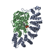 1n4qS S: Starting model for refinement C: citing same article ( |
|---|---|
| Similar structure data |


-Links
 PDBj
PDBj

- Assembly
| Deposited unit | 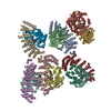
| |||||||||
|---|---|---|---|---|---|---|---|---|---|---|
| 1 | 
| |||||||||
| 2 | 
| |||||||||
| 3 | 
| |||||||||
| 4 | 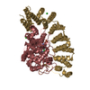
| |||||||||
| 5 | 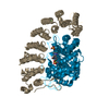
| |||||||||
| 6 | 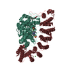
| |||||||||
| Unit cell |
| |||||||||
| Components on special symmetry positions |
|
-Components
-Protein , 2 types, 12 molecules ACEGIKBDFHJL
| #1: Protein | Mass: 44098.145 Da / Num. of mol.: 6 / Fragment: alpha subunit Source method: isolated from a genetically manipulated source Source: (gene. exp.)   Spodoptera frugiperda (fall armyworm) Spodoptera frugiperda (fall armyworm)References: UniProt: Q04631, protein farnesyltransferase, protein geranylgeranyltransferase type I #2: Protein | Mass: 42466.176 Da / Num. of mol.: 6 / Fragment: beta subunit Source method: isolated from a genetically manipulated source Source: (gene. exp.) Spodoptera frugiperda (fall armyworm)References: UniProt: P53610, protein geranylgeranyltransferase type I |
|---|
-Non-polymers , 6 types, 1408 molecules 


| #3: Chemical | ChemComp-ZN /  Mass: 65.409 Da / Num. of mol.: 6 / Source method: obtained synthetically / Formula: Zn Mass: 65.409 Da / Num. of mol.: 6 / Source method: obtained synthetically / Formula: Zn#4: Chemical | ChemComp-SO4 /  Mass: 96.063 Da / Num. of mol.: 6 / Source method: obtained synthetically / Formula: SO4 Mass: 96.063 Da / Num. of mol.: 6 / Source method: obtained synthetically / Formula: SO4#5: Chemical | ChemComp-778 /  Mass: 405.880 Da / Num. of mol.: 6 / Source method: obtained synthetically / Formula: C22H20ClN5O Mass: 405.880 Da / Num. of mol.: 6 / Source method: obtained synthetically / Formula: C22H20ClN5O#6: Chemical | ChemComp-MES /  Mass: 195.237 Da / Num. of mol.: 6 / Source method: obtained synthetically / Formula: C6H13NO4S / Comment: pH buffer*YM Mass: 195.237 Da / Num. of mol.: 6 / Source method: obtained synthetically / Formula: C6H13NO4S / Comment: pH buffer*YM#7: Chemical | ChemComp-CL /  Mass: 35.453 Da / Num. of mol.: 9 / Source method: obtained synthetically / Formula: Cl Mass: 35.453 Da / Num. of mol.: 9 / Source method: obtained synthetically / Formula: Cl#8: Water | ChemComp-HOH / | Mass: 18.015 Da / Num. of mol.: 1375 / Source method: isolated from a natural source / Formula: H2O |
|---|
-Experimental details
-Experiment
| Experiment | Method: X-RAY DIFFRACTION / Number of used crystals: 1 |
|---|
- Sample preparation
| Crystal | Density Matthews: 4.84 Å3/Da / Density % sol: 74.6 % |
|---|---|
| Crystal grow | Temperature: 290 K / pH: 6.3 Details: 1.3 M ammonium sulfate, 175 mM NaCitrate pH 6.5, 100 mM MES pH 6.3, 20 mM DTT, 1uM ZnCl2, VAPOR DIFFUSION, HANGING DROP, temperature 290K, pH 6.30 |
-Data collection
| Diffraction | Mean temperature: 100 K |
|---|---|
| Diffraction source | Source: SYNCHROTRON / Site: APS  / Beamline: 14-BM-C / Wavelength: 1 / Beamline: 14-BM-C / Wavelength: 1 |
| Detector | Type: ADSC QUANTUM 4 / Detector: CCD / Date: Sep 19, 2001 / Details: BENT CONICAL SI-MIRROR (RH COATING) |
| Radiation | Monochromator: BENT GE(III) MONOCHROMATOR / Protocol: SINGLE WAVELENGTH / Monochromatic (M) / Laue (L): M / Scattering type: x-ray |
| Radiation wavelength | Wavelength: 1 Å / Relative weight: 1 |
| Reflection | Resolution: 2.55→30 Å / Num. all: 320017 / Num. obs: 300151 / % possible obs: 93.3 % / Observed criterion σ(I): 0 / Biso Wilson estimate: 38.7 Å2 / Rsym value: 0.05 / Net I/σ(I): 15.2 |
| Reflection shell | Resolution: 2.55→2.64 Å / Mean I/σ(I) obs: 3.1 / Rsym value: 0.256 / % possible all: 85.6 |
- Processing
| Software |
| ||||||||||||||||||||||||||||||||||||||||||||||||||||||||||||||||||||||||||||||||
|---|---|---|---|---|---|---|---|---|---|---|---|---|---|---|---|---|---|---|---|---|---|---|---|---|---|---|---|---|---|---|---|---|---|---|---|---|---|---|---|---|---|---|---|---|---|---|---|---|---|---|---|---|---|---|---|---|---|---|---|---|---|---|---|---|---|---|---|---|---|---|---|---|---|---|---|---|---|---|---|---|---|
| Refinement | Method to determine structure: MOLECULAR REPLACEMENT Starting model: 1N4Q Resolution: 2.55→30 Å / Rfactor Rfree error: 0.002 / Data cutoff high absF: 4876426.32 / Data cutoff low absF: 0 / Isotropic thermal model: RESTRAINED / Cross valid method: THROUGHOUT / σ(F): 0 / Stereochemistry target values: ENGH & HUBER
| ||||||||||||||||||||||||||||||||||||||||||||||||||||||||||||||||||||||||||||||||
| Solvent computation | Solvent model: FLAT MODEL / Bsol: 48.3673 Å2 / ksol: 0.359785 e/Å3 | ||||||||||||||||||||||||||||||||||||||||||||||||||||||||||||||||||||||||||||||||
| Displacement parameters | Biso mean: 57 Å2
| ||||||||||||||||||||||||||||||||||||||||||||||||||||||||||||||||||||||||||||||||
| Refine analyze |
| ||||||||||||||||||||||||||||||||||||||||||||||||||||||||||||||||||||||||||||||||
| Refinement step | Cycle: LAST / Resolution: 2.55→30 Å
| ||||||||||||||||||||||||||||||||||||||||||||||||||||||||||||||||||||||||||||||||
| Refine LS restraints |
| ||||||||||||||||||||||||||||||||||||||||||||||||||||||||||||||||||||||||||||||||
| LS refinement shell | Resolution: 2.55→2.71 Å / Rfactor Rfree error: 0.007 / Total num. of bins used: 6
| ||||||||||||||||||||||||||||||||||||||||||||||||||||||||||||||||||||||||||||||||
| Xplor file |
|
