 Movie
Movie Controller

[English] 日本語
 Yorodumi
- PDB-3mdn: Structure of glutamine aminotransferase class-II domain protein (... -
+ Open data

- Basic information
| Entry | Database: PDB / ID: 3mdn | ||||||
|---|---|---|---|---|---|---|---|
| Title | Structure of glutamine aminotransferase class-II domain protein (SPO2029) from silicibacter pomeroyi | ||||||
 Components Components | Glutamine aminotransferase class-II domain protein | ||||||
 Keywords Keywords | TRANSFERASE / structural genomics / PSI-2 / Protein Structure Initiative / New York SGX Research Center for Structural Genomics / NYSGXRC | ||||||
| Function / homology |  Function and homology information Function and homology information: / Gamma-glutamyl-hercynylcysteine sulfoxide hydrolase EgtC-like / Glutamine amidotransferases class-II / Glutamine amidotransferase type 2 domain profile. / Glutamine amidotransferase type 2 domain / Aminohydrolase, N-terminal nucleophile (Ntn) domain / Glutamine Phosphoribosylpyrophosphate, subunit 1, domain 1 / Nucleophile aminohydrolases, N-terminal / 4-Layer Sandwich / Alpha Beta Similarity search - Domain/homology | ||||||
| Biological species |  Ruegeria pomeroyi (bacteria) Ruegeria pomeroyi (bacteria) | ||||||
| Method |  X-RAY DIFFRACTION / SYNCHROTRON / SAD / Resolution: 2.09 Å X-RAY DIFFRACTION / SYNCHROTRON / SAD / Resolution: 2.09 Å | ||||||
 Authors Authors | Ramagopal, U.A. / Toro, R. / Burley, S.K. / Almo, S.C. / New York SGX Research Center for Structural Genomics (NYSGXRC) | ||||||
 Citation Citation | Journal: To be published Title: Structure of glutamine aminotransferase class-II domain protein (SPO2029) from silicibacter pomeroyi Authors: Ramagopal, U.A. / Toro, R. / Burley, S.K. / Almo, S.C. | ||||||
| History |
|
- Structure visualization
| Structure viewer | Molecule: MolmilJmol/JSmol |
|---|
- Downloads & links
-Download
| PDBx/mmCIF format | 3mdn.cif.gz | 180.6 KB | Display | PDBx/mmCIF format |
|---|---|---|---|---|
| PDB format | pdb3mdn.ent.gz | 144 KB | Display | PDB format |
| PDBx/mmJSON format | 3mdn.json.gz | Tree view | PDBx/mmJSON format | |
| Others |  Other downloads Other downloads |
-Validation report
| Summary document | 3mdn_validation.pdf.gz | 462.3 KB | Display | wwPDB validaton report |
|---|---|---|---|---|
| Full document | 3mdn_full_validation.pdf.gz | 482.6 KB | Display | |
| Data in XML | 3mdn_validation.xml.gz | 32.4 KB | Display | |
| Data in CIF | 3mdn_validation.cif.gz | 44 KB | Display | |
| Arichive directory | https://data.pdbj.org/pub/pdb/validation_reports/md/3mdnftp://data.pdbj.org/pub/pdb/validation_reports/md/3mdn | HTTPS FTP |
-Related structure data
| Similar structure data | |
|---|---|
| Other databases |


-Links
 PDBj
PDBj


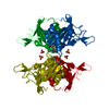
- Assembly
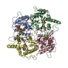
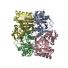
| Deposited unit | 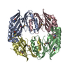
| ||||||||
|---|---|---|---|---|---|---|---|---|---|
| 1 |
| ||||||||
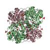
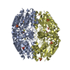
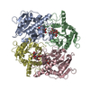
| Unit cell |
|
-Components
| #1: Protein | Mass: 30600.229 Da / Num. of mol.: 4 Source method: isolated from a genetically manipulated source Source: (gene. exp.) Ruegeria pomeroyi (bacteria) / Gene: SPO2029 / Plasmid: BC-pSGX4(BC) / Production host: #2: Water | ChemComp-HOH / |  Mass: 18.015 Da / Num. of mol.: 36 / Source method: isolated from a natural source / Formula: H2O Mass: 18.015 Da / Num. of mol.: 36 / Source method: isolated from a natural source / Formula: H2OHas protein modification | Y | |
|---|
-Experimental details
-Experiment
| Experiment | Method: X-RAY DIFFRACTION / Number of used crystals: 1 |
|---|
- Sample preparation
| Crystal | Density Matthews: 2.11 Å3/Da / Density % sol: 41.7 % |
|---|---|
| Crystal grow | Temperature: 298 K / Method: vapor diffusion, sitting drop / pH: 7.5 Details: 0.2M MgCl2, 25% PEG 3350, pH 7.5, Vapor diffusion, Sitting drop, temperature 298K |
-Data collection
| Diffraction | Mean temperature: 100 K | |||||||||||||||||||||||||||||||||||||||||||||||||||||||||||||||||||||||||||||||||||||||||||||||||||||||||||||||||||||||||||||||||||||||||||||||||||
|---|---|---|---|---|---|---|---|---|---|---|---|---|---|---|---|---|---|---|---|---|---|---|---|---|---|---|---|---|---|---|---|---|---|---|---|---|---|---|---|---|---|---|---|---|---|---|---|---|---|---|---|---|---|---|---|---|---|---|---|---|---|---|---|---|---|---|---|---|---|---|---|---|---|---|---|---|---|---|---|---|---|---|---|---|---|---|---|---|---|---|---|---|---|---|---|---|---|---|---|---|---|---|---|---|---|---|---|---|---|---|---|---|---|---|---|---|---|---|---|---|---|---|---|---|---|---|---|---|---|---|---|---|---|---|---|---|---|---|---|---|---|---|---|---|---|---|---|---|
| Diffraction source | Source: SYNCHROTRON / Site: NSLS  / Beamline: X29A / Wavelength: 0.9793 Å / Beamline: X29A / Wavelength: 0.9793 Å | |||||||||||||||||||||||||||||||||||||||||||||||||||||||||||||||||||||||||||||||||||||||||||||||||||||||||||||||||||||||||||||||||||||||||||||||||||
| Detector | Type: ADSC QUANTUM 315 / Detector: CCD / Date: Feb 2, 2010 | |||||||||||||||||||||||||||||||||||||||||||||||||||||||||||||||||||||||||||||||||||||||||||||||||||||||||||||||||||||||||||||||||||||||||||||||||||
| Radiation | Protocol: SINGLE WAVELENGTH / Scattering type: x-ray | |||||||||||||||||||||||||||||||||||||||||||||||||||||||||||||||||||||||||||||||||||||||||||||||||||||||||||||||||||||||||||||||||||||||||||||||||||
| Radiation wavelength | Wavelength: 0.9793 Å / Relative weight: 1 | |||||||||||||||||||||||||||||||||||||||||||||||||||||||||||||||||||||||||||||||||||||||||||||||||||||||||||||||||||||||||||||||||||||||||||||||||||
| Reflection | Resolution: 2.09→40 Å / Num. obs: 59569 / % possible obs: 99.8 % / Redundancy: 6.1 % / Rmerge(I) obs: 0.063 / Χ2: 1.283 / Net I/σ(I): 13.4 | |||||||||||||||||||||||||||||||||||||||||||||||||||||||||||||||||||||||||||||||||||||||||||||||||||||||||||||||||||||||||||||||||||||||||||||||||||
| Reflection shell |
|
- Processing
| Software |
| |||||||||||||||||||||||||||||||||||||||||||||||||||||||||||||||||||||||||||||||||||||||||||||||||||||||||||||||||||||||||||||
|---|---|---|---|---|---|---|---|---|---|---|---|---|---|---|---|---|---|---|---|---|---|---|---|---|---|---|---|---|---|---|---|---|---|---|---|---|---|---|---|---|---|---|---|---|---|---|---|---|---|---|---|---|---|---|---|---|---|---|---|---|---|---|---|---|---|---|---|---|---|---|---|---|---|---|---|---|---|---|---|---|---|---|---|---|---|---|---|---|---|---|---|---|---|---|---|---|---|---|---|---|---|---|---|---|---|---|---|---|---|---|---|---|---|---|---|---|---|---|---|---|---|---|---|---|---|---|
| Refinement | Method to determine structure: SAD / Resolution: 2.09→40 Å / Cor.coef. Fo:Fc: 0.962 / Cor.coef. Fo:Fc free: 0.94 / WRfactor Rfree: 0.27 / WRfactor Rwork: 0.224 / Occupancy max: 1 / Occupancy min: 1 / FOM work R set: 0.796 / SU B: 12.584 / SU ML: 0.148 / SU R Cruickshank DPI: 0.213 / SU Rfree: 0.189 / TLS residual ADP flag: LIKELY RESIDUAL / Cross valid method: THROUGHOUT / σ(F): 0 / ESU R: 0.213 / ESU R Free: 0.188 / Stereochemistry target values: MAXIMUM LIKELIHOOD Details: HYDROGENS HAVE BEEN ADDED IN THE RIDING POSITIONS U VALUES : RESIDUAL ONLY
| |||||||||||||||||||||||||||||||||||||||||||||||||||||||||||||||||||||||||||||||||||||||||||||||||||||||||||||||||||||||||||||
| Solvent computation | Ion probe radii: 0.8 Å / Shrinkage radii: 0.8 Å / VDW probe radii: 1.4 Å / Solvent model: MASK | |||||||||||||||||||||||||||||||||||||||||||||||||||||||||||||||||||||||||||||||||||||||||||||||||||||||||||||||||||||||||||||
| Displacement parameters | Biso max: 59.43 Å2 / Biso mean: 31.206 Å2 / Biso min: 11.81 Å2
| |||||||||||||||||||||||||||||||||||||||||||||||||||||||||||||||||||||||||||||||||||||||||||||||||||||||||||||||||||||||||||||
| Refinement step | Cycle: LAST / Resolution: 2.09→40 Å
| |||||||||||||||||||||||||||||||||||||||||||||||||||||||||||||||||||||||||||||||||||||||||||||||||||||||||||||||||||||||||||||
| Refine LS restraints |
| |||||||||||||||||||||||||||||||||||||||||||||||||||||||||||||||||||||||||||||||||||||||||||||||||||||||||||||||||||||||||||||
| LS refinement shell | Resolution: 2.095→2.149 Å / Total num. of bins used: 20
| |||||||||||||||||||||||||||||||||||||||||||||||||||||||||||||||||||||||||||||||||||||||||||||||||||||||||||||||||||||||||||||
| Refinement TLS params. | Method: refined / Refine-ID: X-RAY DIFFRACTION
| |||||||||||||||||||||||||||||||||||||||||||||||||||||||||||||||||||||||||||||||||||||||||||||||||||||||||||||||||||||||||||||
| Refinement TLS group |
|
