Component-ID: 1 / Ens-ID: 1 / Beg auth comp-ID: MSE / Beg label comp-ID: MSE / End auth comp-ID: LEU / End label comp-ID: LEU / Refine code: 1 / Auth seq-ID: 1 - 217 / Label seq-ID: 2 - 218
Dom-ID
Auth asym-ID
Label asym-ID
1
A
A
2
B
B
Details
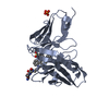
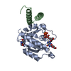
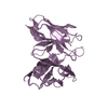
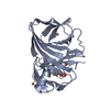
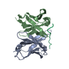
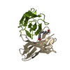
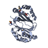
CRYSTAL PACKING ANALYSIS AND ANALYTICAL SIZE-EXCLUSION CHROMATOGRAPHY SUPPORT THE ASSIGNMENT OF A MONOMER AS THE SIGNIFICANT OLIGOMERIC FORM IN SOLUTION.
-
Components
#1: Protein
Putativemonooxygenase
Mass: 25422.156 Da / Num. of mol.: 2 Source method: isolated from a genetically manipulated source Source: (gene. exp.) Fusobacterium nucleatum subsp. nucleatum (bacteria) Strain: ATCC 25586 / Gene: FN1347 / Plasmid: SpeedET / Production host: Escherichia Coli (E. coli) / Strain (production host): HK100 / References: UniProt: Q8RDZ4
Mass: 18.015 Da / Num. of mol.: 41 / Source method: isolated from a natural source / Formula: H2O
Has protein modification
Y
Sequence details
THE CONSTRUCT WAS EXPRESSED WITH A PURIFICATION TAG MGSDKIHHHHHHENLYFQG. THE TAG WAS REMOVED WITH ...THE CONSTRUCT WAS EXPRESSED WITH A PURIFICATION TAG MGSDKIHHHHHHENLYFQG. THE TAG WAS REMOVED WITH TEV PROTEASE LEAVING ONLY A GLYCINE (0) FOLLOWED BY THE TARGET SEQUENCE.
-
Experimental details
-
Experiment
Experiment
Method: X-RAY DIFFRACTION / Number of used crystals: 1
-
Sample preparation
Crystal
Density Matthews: 2.23 Å3/Da / Density % sol: 44.86 %
Crystal grow
Temperature: 277 K / Method: vapor diffusion, sitting drop / pH: 5.6 Details: 0.2000M NH4OAc, 30.0000% PEG-4000, 0.1M Citrate pH 5.6, NANODROP, VAPOR DIFFUSION, SITTING DROP, temperature 277K
Type: MARMOSAIC 325 mm CCD / Detector: CCD / Date: Dec 3, 2009 / Details: Flat mirror (vertical focusing)
Radiation
Monochromator: Single crystal Si(111) bent monochromator (horizontal focusing) Protocol: MAD / Monochromatic (M) / Laue (L): M / Scattering type: x-ray
Radiation wavelength
ID
Wavelength (Å)
Relative weight
1
0.91837
1
2
0.97925
1
3
0.97894
1
Reflection
Resolution: 2.55→29.607 Å / Num. obs: 14638 / % possible obs: 97.9 % / Observed criterion σ(I): -3 / Biso Wilson estimate: 53.814 Å2 / Rmerge(I) obs: 0.078 / Net I/σ(I): 9
Reflection shell
Resolution (Å)
Rmerge(I) obs
Mean I/σ(I) obs
Num. measured obs
Num. unique obs
Diffraction-ID
% possible all
2.55-2.64
0.654
1.3
5184
2693
1
95.7
2.64-2.75
0.457
1.9
5705
2946
1
99
2.75-2.87
0.333
2.6
5333
2740
1
98.6
2.87-3.02
0.241
3.5
5515
2838
1
99
3.02-3.21
0.168
5.1
5480
2819
1
98.5
3.21-3.46
0.101
7.9
5529
2834
1
98.1
3.46-3.8
0.07
11.3
5315
2727
1
97.8
3.8-4.35
0.05
15
5511
2817
1
97.4
4.35-5.46
0.036
19.1
5454
2783
1
97.7
5.46-29.607
0.028
22
5634
2875
1
97.7
-
Phasing
Phasing
Method: MAD
-
Processing
Software
Name
Version
Classification
NB
REFMAC
5.5.0102
refinement
PHENIX
refinement
SHELX
phasing
MolProbity
3beta29
modelbuilding
XSCALE
datascaling
PDB_EXTRACT
3.006
dataextraction
XDS
datareduction
SHELXD
phasing
autoSHARP
phasing
Refinement
Method to determine structure: MAD / Resolution: 2.55→29.607 Å / Cor.coef. Fo:Fc: 0.937 / Cor.coef. Fo:Fc free: 0.904 / Occupancy max: 1 / Occupancy min: 0.5 / SU B: 30.986 / SU ML: 0.301 / TLS residual ADP flag: LIKELY RESIDUAL / Cross valid method: THROUGHOUT / σ(F): 0 / ESU R Free: 0.358 Stereochemistry target values: MAXIMUM LIKELIHOOD WITH PHASES Details: 1.HYDROGENS HAVE BEEN ADDED IN THE RIDING POSITIONS.2.A MET-INHIBITION PROTOCOL WAS USED FOR SELENOMETHIONIN INCORPORATION DURING PROTEIN EXPRESSION. THE OCCUPANCY OF THE SE ATOMS IN THE MSE ...Details: 1.HYDROGENS HAVE BEEN ADDED IN THE RIDING POSITIONS.2.A MET-INHIBITION PROTOCOL WAS USED FOR SELENOMETHIONIN INCORPORATION DURING PROTEIN EXPRESSION. THE OCCUPANCY OF THE SE ATOMS IN THE MSE RESIDUES WAS REDUCED TO 0.75 TO ACCOUNT FOR THE REDUCED SCATTERING POWER DUE TO PARTIAL S-MET INCORPORATION. 3 .ACETATE (ACT) FROM THE CRYSTALLIZATION WAS MODELED INTO THE STRUCTURE 4.ATOM RECORD CONTAINS RESIDUAL B FACTORS ONLY. 4. ASN 90 ON BOTH THE A AND B CHAINS ARE FLAGGED AS MOLPROBITY RAMACHANDRAN OUTLIERS, AND IT IS LIKELY THAT THIS IS RELATED TO DISORDERED ELECTRON DENSITIES AT THIS RESIDUE.
Rfactor
Num. reflection
% reflection
Selection details
Rfree
0.261
1470
10.1 %
RANDOM
Rwork
0.212
-
-
-
obs
0.217
14626
99.44 %
-
Solvent computation
Ion probe radii: 0.8 Å / Shrinkage radii: 0.8 Å / VDW probe radii: 1.4 Å / Solvent model: BABINET MODEL WITH MASK
In the structure databanks used in Yorodumi, some data are registered as the other names, "COVID-19 virus" and "2019-nCoV". Here are the details of the virus and the list of structure data.
Jan 31, 2019. EMDB accession codes are about to change! (news from PDBe EMDB page)
EMDB accession codes are about to change! (news from PDBe EMDB page)
The allocation of 4 digits for EMDB accession codes will soon come to an end. Whilst these codes will remain in use, new EMDB accession codes will include an additional digit and will expand incrementally as the available range of codes is exhausted. The current 4-digit format prefixed with “EMD-” (i.e. EMD-XXXX) will advance to a 5-digit format (i.e. EMD-XXXXX), and so on. It is currently estimated that the 4-digit codes will be depleted around Spring 2019, at which point the 5-digit format will come into force.
The EM Navigator/Yorodumi systems omit the EMD- prefix.
Related info.:Q: What is EMD? / ID/Accession-code notation in Yorodumi/EM Navigator
Yorodumi is a browser for structure data from EMDB, PDB, SASBDB, etc.
This page is also the successor to EM Navigator detail page, and also detail information page/front-end page for Omokage search.
The word "yorodu" (or yorozu) is an old Japanese word meaning "ten thousand". "mi" (miru) is to see.
Related info.:EMDB / PDB / SASBDB / Comparison of 3 databanks / Yorodumi Search / Aug 31, 2016. New EM Navigator & Yorodumi / Yorodumi Papers / Jmol/JSmol / Function and homology information / Changes in new EM Navigator and Yorodumi
 Movie
Movie Controller

 Yorodumi
 Open data
 Basic information
 Components
 Keywords
 Function and homology information
 Fusobacterium nucleatum subsp. nucleatum (bacteria)
 X-RAY DIFFRACTION /
  Authors
 Citation
 Structure visualization
 Downloads & links
 Other downloads
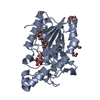


 PDBj
PDBj Assembly

 Mass: 59.044 Da / Num. of mol.: 2 / Source method: obtained synthetically / Formula: C2H3O2
 Mass: 18.015 Da / Num. of mol.: 41 / Source method: isolated from a natural source / Formula: H2O
 Sample preparation
 / Beamline: BL11-1 / Wavelength: 0.91837,0.97925,0.97894
 Processing