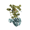
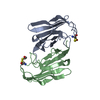
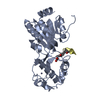
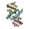
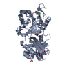
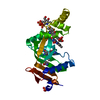
[English] 日本語
Yorodumi
- PDB-3m7f: Crystal structure of the Nedd4 C2/Grb10 SH2 complex -

+
Open data


ID or keywords:

Loading...

-
Basic information

Entry
Database: PDB / ID: 3m7f
TitleCrystal structure of the Nedd4 C2/Grb10 SH2 complex
Components
  • E3 ubiquitin-protein ligase NEDD4
  • Growth factor receptor-bound protein 10
KeywordsSIGNALING PROTEIN/LIGASE / Nedd4 / C2 domain / Grb10 / SH2 domain / Phosphoprotein / Ligase / Ubl conjugation pathway / SIGNALING PROTEIN-LIGASE complex
Function / homology
Function and homology information


formation of structure involved in a symbiotic process / FLT3 Signaling / positive regulation of nucleocytoplasmic transport / Regulation of PTEN localization / Downregulation of ERBB4 signaling / IRS activation / Insulin receptor signalling cascade / ISG15 antiviral mechanism / Signal attenuation / Signaling by SCF-KIT ...formation of structure involved in a symbiotic process / FLT3 Signaling / positive regulation of nucleocytoplasmic transport / Regulation of PTEN localization / Downregulation of ERBB4 signaling / IRS activation / Insulin receptor signalling cascade / ISG15 antiviral mechanism / Signal attenuation / Signaling by SCF-KIT / vascular associated smooth muscle cell migration / Regulation of PTEN stability and activity / negative regulation of sodium ion transport / nuclear receptor-mediated glucocorticoid signaling pathway / viral budding / negative regulation of potassium ion export across plasma membrane / endocardial cushion development / negative regulation of glycogen biosynthetic process / phosphothreonine residue binding / negative regulation of D-glucose import / RET signaling / receptor catabolic process / protein targeting to lysosome / potassium channel inhibitor activity / ubiquitin-dependent protein catabolic process via the multivesicular body sorting pathway / HECT-type E3 ubiquitin transferase / Antigen processing: Ubiquitination & Proteasome degradation / sodium channel inhibitor activity / proline-rich region binding / positive regulation of vascular endothelial growth factor receptor signaling pathway / RNA polymerase binding / blood vessel morphogenesis / beta-2 adrenergic receptor binding / regulation of dendrite morphogenesis / negative regulation of vascular endothelial growth factor receptor signaling pathway / sodium ion transport / neuromuscular junction development / regulation of synapse organization / negative regulation of Wnt signaling pathway / outflow tract morphogenesis / protein K63-linked ubiquitination / phosphoserine residue binding / protein monoubiquitination / ubiquitin ligase complex / postsynaptic cytosol / progesterone receptor signaling pathway / ERK1 and ERK2 cascade / phosphotyrosine residue binding / ionotropic glutamate receptor binding / negative regulation of insulin receptor signaling pathway / insulin-like growth factor receptor signaling pathway / T cell activation / ubiquitin binding / regulation of membrane potential / insulin receptor binding / establishment of localization in cell / receptor internalization / neuron projection development / positive regulation of protein catabolic process / cellular response to UV / ubiquitin protein ligase activity / signaling receptor complex adaptor activity / positive regulation of cold-induced thermogenesis / ubiquitin-dependent protein catabolic process / cell cortex / protein-macromolecule adaptor activity / adaptive immune response / gene expression / transmembrane transporter binding / positive regulation of phosphatidylinositol 3-kinase/protein kinase B signal transduction / protein ubiquitination / protein domain specific binding / innate immune response / DNA damage response / chromatin / perinuclear region of cytoplasm / glutamatergic synapse / negative regulation of transcription by RNA polymerase II / Golgi apparatus / signal transduction / protein-containing complex / nucleoplasm / nucleus / membrane / plasma membrane / cytosol / cytoplasm
Similarity search - Function
Grb10, SH2 / BPS (Between PH and SH2) domain / BPS (Between PH and SH2) / GRB/APBB1IP / APBB1IP, PH domain / RA like domain / Ras association (RalGDS/AF-6) domain / E3 ubiquitin-protein ligase, SMURF1 type / Ras-associating (RA) domain profile. / Ras-associating (RA) domain ...Grb10, SH2 / BPS (Between PH and SH2) domain / BPS (Between PH and SH2) / GRB/APBB1IP / APBB1IP, PH domain / RA like domain / Ras association (RalGDS/AF-6) domain / E3 ubiquitin-protein ligase, SMURF1 type / Ras-associating (RA) domain profile. / Ras-associating (RA) domain / : / HECT domain / HECT, E3 ligase catalytic domain / HECT-domain (ubiquitin-transferase) / HECT domain profile. / Domain Homologous to E6-AP Carboxyl Terminus with / C2 domain / SH2 domain / SHC Adaptor Protein / Protein kinase C conserved region 2 (CalB) / C2 domain / WW domain / WW/rsp5/WWP domain signature. / C2 domain / C2 domain profile. / WW domain superfamily / WW/rsp5/WWP domain profile. / Domain with 2 conserved Trp (W) residues / WW domain / PH domain / C2 domain superfamily / PH domain profile. / Pleckstrin homology domain. / Pleckstrin homology domain / SH2 domain / Src homology 2 (SH2) domain profile. / Src homology 2 domains / SH2 domain / SH2 domain superfamily / PH-like domain superfamily / Ubiquitin-like domain superfamily / Immunoglobulin-like / Sandwich / 2-Layer Sandwich / Mainly Beta / Alpha Beta
Similarity search - Domain/homology
E3 ubiquitin-protein ligase NEDD4 / Growth factor receptor-bound protein 10
Similarity search - Component
Biological speciesMus musculus (house mouse)
MethodX-RAY DIFFRACTION / SYNCHROTRON / MOLECULAR REPLACEMENT / Resolution: 2 Å
AuthorsHuang, Q. / Szebenyi, M.
CitationJournal: J.Biol.Chem. / Year: 2010
Title: Structural Basis for the Interaction between the Growth Factor-binding Protein GRB10 and the E3 Ubiquitin Ligase NEDD4.
Authors: Huang, Q. / Szebenyi, D.M.
History
DepositionMar 16, 2010Deposition site: RCSB / Processing site: RCSB
Revision 1.0Oct 27, 2010Provider: repository / Type: Initial release
Revision 1.1Jul 13, 2011Group: Version format compliance
Revision 1.2Sep 6, 2023Group: Data collection / Database references / Refinement description
Category: chem_comp_atom / chem_comp_bond ...chem_comp_atom / chem_comp_bond / database_2 / pdbx_initial_refinement_model
Item: _database_2.pdbx_DOI / _database_2.pdbx_database_accession

-
Structure visualization

Structure viewerMolecule:
MolmilJmol/JSmol

Downloads & links

-
Assembly

Deposited unit
A: Growth factor receptor-bound protein 10
B: E3 ubiquitin-protein ligase NEDD4


Theoretical massNumber of molelcules
Total (without water)32,9842
Polymers32,9842
Non-polymers00
Water2,540141
1


  • Idetical with deposited unit
  • defined by author&software
TypeNameSymmetry operationNumber
identity operation1_555x,y,z1
Buried area1890 Å2
ΔGint-4 kcal/mol
Surface area13350 Å2
MethodPISA
Unit cell
Length a, b, c (Å)52.163, 70.086, 86.246
Angle α, β, γ (deg.)90.00, 90.00, 90.00
Int Tables number19
Space group name H-MP212121

-
Components

#1: Protein Growth factor receptor-bound protein 10 / GRB10 adapter protein / Maternally expressed gene 1 protein


Mass: 12644.569 Da / Num. of mol.: 1 / Fragment: UNP residues 514-621, SH2 domain
Source method: isolated from a genetically manipulated source
Source: (gene. exp.) Mus musculus (house mouse) / Gene: Grb10, Kiaa0093, Meg1, Nedd-4, Nedd4, Nedd4a / Production host: Escherichia coli (E. coli) / Strain (production host): BL21(DE3)
References: UniProt: Q60760, Ligases; Forming carbon-nitrogen bonds; Acid-amino-acid ligases (peptide synthases)
#2: Protein E3 ubiquitin-protein ligase NEDD4 / Neural precursor cell expressed developmentally down-regulated protein 4 / NEDD-4


Mass: 20339.244 Da / Num. of mol.: 1 / Fragment: UNP residues 71-246, C2 domain
Source method: isolated from a genetically manipulated source
Source: (gene. exp.) Mus musculus (house mouse) / Gene: Grb10, Kiaa0093, Meg1, Nedd-4, Nedd4, Nedd4a / Production host: Escherichia coli (E. coli) / Strain (production host): BL21(DE3)
References: UniProt: P46935, Ligases; Forming carbon-nitrogen bonds; Acid-amino-acid ligases (peptide synthases)
#3: Water ChemComp-HOH / water


Mass: 18.015 Da / Num. of mol.: 141 / Source method: isolated from a natural source / Formula: H2O
Sequence detailsTHIS RESIDUE AT POSITION 583 WAS IDENTIFIED AS SER BASED ON THE ELECTRON DENSITY

-
Experimental details

-
Experiment

ExperimentMethod: X-RAY DIFFRACTION / Number of used crystals: 1

-
Sample preparation

CrystalDensity Matthews: 2.39 Å3/Da / Density % sol: 48.53 %
Crystal growTemperature: 295 K / Method: evaporation / pH: 6.5
Details: crystallization in 35% MPD, pH 6.5, EVAPORATION, temperature 295K

-
Data collection

DiffractionMean temperature: 100 K
Diffraction sourceSource: SYNCHROTRON / Site: CHESS / Beamline: A1 / Wavelength: 0.978 Å
DetectorType: ADSC QUANTUM 210 / Detector: CCD / Date: Oct 28, 2009 / Details: mirrors
RadiationMonochromator: mirrors / Protocol: SINGLE WAVELENGTH / Monochromatic (M) / Laue (L): M / Scattering type: x-ray
Radiation wavelengthWavelength: 0.978 Å / Relative weight: 1
ReflectionResolution: 2→50 Å / Num. obs: 21866 / Redundancy: 6.5 % / Rmerge(I) obs: 0.052

-
Processing

Software
NameVersionClassification
HKL-2000data collection
PHASERphasing
PHENIX(phenix.refine: 1.6_289)refinement
HKL-2000data reduction
HKL-2000data scaling
RefinementMethod to determine structure: MOLECULAR REPLACEMENT
Starting model: PDB ENTRIES 1NRV AND 3B7Y
Resolution: 2→43.123 Å / SU ML: 0.25 / σ(F): 0.06 / Stereochemistry target values: ML
RfactorNum. reflection% reflection
Rfree0.232 1081 5.15 %
Rwork0.1925 --
obs0.1945 20973 95.58 %
Solvent computationShrinkage radii: 0.9 Å / VDW probe radii: 1.11 Å / Solvent model: FLAT BULK SOLVENT MODEL / Bsol: 53.194 Å2 / ksol: 0.359 e/Å3
Refine analyzeLuzzati sigma a obs: 0.25 Å
Refinement stepCycle: LAST / Resolution: 2→43.123 Å
ProteinNucleic acidLigandSolventTotal
Num. atoms1985 0 0 141 2126
Refine LS restraints
Refine-IDTypeDev idealNumber
X-RAY DIFFRACTIONf_bond_d0.0062055
X-RAY DIFFRACTIONf_angle_d1.0572772
X-RAY DIFFRACTIONf_dihedral_angle_d17.612759
X-RAY DIFFRACTIONf_chiral_restr0.077305
X-RAY DIFFRACTIONf_plane_restr0.004358
LS refinement shell
Resolution (Å)Rfactor RfreeNum. reflection RfreeRfactor RworkNum. reflection RworkRefine-ID% reflection obs (%)
2-2.09270.23951290.20472163X-RAY DIFFRACTION85
2.0927-2.20310.27911210.1922346X-RAY DIFFRACTION92
2.2031-2.34110.26221330.20022429X-RAY DIFFRACTION95
2.3411-2.52180.29171260.20542481X-RAY DIFFRACTION97
2.5218-2.77560.26611360.20622547X-RAY DIFFRACTION98
2.7756-3.17710.24381510.20482575X-RAY DIFFRACTION99
3.1771-4.00240.21371270.16712647X-RAY DIFFRACTION100
4.0024-43.1330.19861580.18622704X-RAY DIFFRACTION99
Refinement TLS params.Method: refined / Origin x: 6.5699 Å / Origin y: 5.1861 Å / Origin z: 6.4036 Å
111213212223313233
T0.2243 Å20.0074 Å20.0142 Å2-0.2038 Å20.0029 Å2--0.2334 Å2
L1.9337 °2-0.0547 °21.5702 °2-0.8154 °2-0.1361 °2--2.0747 °2
S-0.0099 Å °-0.0891 Å °-0.0584 Å °0.149 Å °-0.0141 Å °0.0529 Å °-0.0321 Å °-0.0148 Å °-0 Å °
Refinement TLS groupSelection details: all

+
About Yorodumi

-
News

-
Feb 9, 2022. New format data for meta-information of EMDB entries

New format data for meta-information of EMDB entries

  • Version 3 of the EMDB header file is now the official format.
  • The previous official version 1.9 will be removed from the archive.

Related info.:EMDB header

External links:wwPDB to switch to version 3 of the EMDB data model

-
Aug 12, 2020. Covid-19 info

Covid-19 info

URL: https://pdbj.org/emnavi/covid19.php

New page: Covid-19 featured information page in EM Navigator.

Related info.:Covid-19 info / Mar 5, 2020. Novel coronavirus structure data

+
Mar 5, 2020. Novel coronavirus structure data

Novel coronavirus structure data

Related info.:Yorodumi Speices / Aug 12, 2020. Covid-19 info

External links:COVID-19 featured content - PDBj / Molecule of the Month (242):Coronavirus Proteases

+
Jan 31, 2019. EMDB accession codes are about to change! (news from PDBe EMDB page)

EMDB accession codes are about to change! (news from PDBe EMDB page)

  • The allocation of 4 digits for EMDB accession codes will soon come to an end. Whilst these codes will remain in use, new EMDB accession codes will include an additional digit and will expand incrementally as the available range of codes is exhausted. The current 4-digit format prefixed with “EMD-” (i.e. EMD-XXXX) will advance to a 5-digit format (i.e. EMD-XXXXX), and so on. It is currently estimated that the 4-digit codes will be depleted around Spring 2019, at which point the 5-digit format will come into force.
  • The EM Navigator/Yorodumi systems omit the EMD- prefix.

Related info.:Q: What is EMD? / ID/Accession-code notation in Yorodumi/EM Navigator

External links:EMDB Accession Codes are Changing Soon! / Contact to PDBj

+
Jul 12, 2017. Major update of PDB

Major update of PDB

  • wwPDB released updated PDB data conforming to the new PDBx/mmCIF dictionary.
  • This is a major update changing the version number from 4 to 5, and with Remediation, in which all the entries are updated.
  • In this update, many items about electron microscopy experimental information are reorganized (e.g. em_software).
  • Now, EM Navigator and Yorodumi are based on the updated data.

External links:wwPDB Remediation / Enriched Model Files Conforming to OneDep Data Standards Now Available in the PDB FTP Archive

-
Yorodumi

Thousand views of thousand structures

  • Yorodumi is a browser for structure data from EMDB, PDB, SASBDB, etc.
  • This page is also the successor to EM Navigator detail page, and also detail information page/front-end page for Omokage search.
  • The word "yorodu" (or yorozu) is an old Japanese word meaning "ten thousand". "mi" (miru) is to see.

Related info.:EMDB / PDB / SASBDB / Comparison of 3 databanks / Yorodumi Search / Aug 31, 2016. New EM Navigator & Yorodumi / Yorodumi Papers / Jmol/JSmol / Function and homology information / Changes in new EM Navigator and Yorodumi

Read more