 Movie
Movie Controller


+ Open data

- Basic information
| Entry | Database: PDB / ID: 1nrv | ||||||
|---|---|---|---|---|---|---|---|
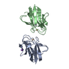
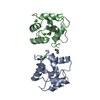
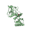
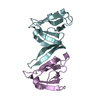
| Title | Crystal structure of the SH2 domain of Grb10 | ||||||
 Components Components | Growth factor receptor-bound protein 10 | ||||||
 Keywords Keywords | SIGNALING PROTEIN / DIMER | ||||||
| Function / homology |  Function and homology information Function and homology informationResponse of EIF2AK1 (HRI) to heme deficiency / negative regulation of glycogen biosynthetic process / negative regulation of D-glucose import across plasma membrane / positive regulation of vascular endothelial growth factor receptor signaling pathway / IRS activation / negative regulation of Wnt signaling pathway / RET signaling / positive regulation of phosphorylation / Signal attenuation / negative regulation of insulin receptor signaling pathway ...Response of EIF2AK1 (HRI) to heme deficiency / negative regulation of glycogen biosynthetic process / negative regulation of D-glucose import across plasma membrane / positive regulation of vascular endothelial growth factor receptor signaling pathway / IRS activation / negative regulation of Wnt signaling pathway / RET signaling / positive regulation of phosphorylation / Signal attenuation / negative regulation of insulin receptor signaling pathway / FLT3 Signaling / Insulin receptor signalling cascade / insulin receptor binding / Signaling by SCF-KIT / response to insulin / insulin receptor signaling pathway / positive regulation of cold-induced thermogenesis / signaling receptor complex adaptor activity / intracellular signal transduction / protein-containing complex / identical protein binding / plasma membrane / cytoplasm / cytosol Similarity search - Function | ||||||
| Biological species |  Homo sapiens (human) Homo sapiens (human) | ||||||
| Method |  X-RAY DIFFRACTION / SYNCHROTRON / MOLECULAR REPLACEMENT / Resolution: 1.65 Å X-RAY DIFFRACTION / SYNCHROTRON / MOLECULAR REPLACEMENT / Resolution: 1.65 Å | ||||||
 Authors Authors | Stein, E.G. / Hubbard, S.R. | ||||||
 Citation Citation | Journal: J.Biol.Chem. / Year: 2003 Title: Structural basis for dimerization of the Grb10 Src homology 2 domain. Implications for ligand specificity. Authors: Stein, E.G. / Ghirlando, R. / Hubbard, S.R. | ||||||
| History |
|
- Structure visualization
| Structure viewer | Molecule: MolmilJmol/JSmol |
|---|
- Downloads & links
-Download
| PDBx/mmCIF format | 1nrv.cif.gz | 56.6 KB | Display | PDBx/mmCIF format |
|---|---|---|---|---|
| PDB format | pdb1nrv.ent.gz | 40.6 KB | Display | PDB format |
| PDBx/mmJSON format | 1nrv.json.gz | Tree view | PDBx/mmJSON format | |
| Others |  Other downloads Other downloads |
-Validation report
| Arichive directory | https://data.pdbj.org/pub/pdb/validation_reports/nr/1nrvftp://data.pdbj.org/pub/pdb/validation_reports/nr/1nrv | HTTPS FTP |
|---|
-Related structure data
| Related structure data | 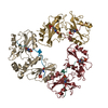 1a81S S: Starting model for refinement |
|---|---|
| Similar structure data |


-Links
 PDBj
PDBj


- Assembly
| Deposited unit | 
| ||||||||
|---|---|---|---|---|---|---|---|---|---|
| 1 |
| ||||||||
| Unit cell |
| ||||||||
| Details | THE BIOLOGICAL ASSEMBLY IS THE DIMER IN THE ASYMMETRIC UNIT. |
-Components
| #1: Protein | Mass: 12361.202 Da / Num. of mol.: 2 / Fragment: SH2 domain Source method: isolated from a genetically manipulated source Source: (gene. exp.) Homo sapiens (human) / Gene: GRB10 OR GRBIR OR KIAA0207 / Plasmid: pET21 / Species (production host): Escherichia coli / Production host:  #2: Water | ChemComp-HOH / |  Mass: 18.015 Da / Num. of mol.: 179 / Source method: isolated from a natural source / Formula: H2O Mass: 18.015 Da / Num. of mol.: 179 / Source method: isolated from a natural source / Formula: H2O |
|---|
-Experimental details
-Experiment
| Experiment | Method: X-RAY DIFFRACTION / Number of used crystals: 1 |
|---|
- Sample preparation
| Crystal | Density Matthews: 2.17 Å3/Da / Density % sol: 42.76 % | |||||||||||||||||||||||||||||||||||||||||||||||||
|---|---|---|---|---|---|---|---|---|---|---|---|---|---|---|---|---|---|---|---|---|---|---|---|---|---|---|---|---|---|---|---|---|---|---|---|---|---|---|---|---|---|---|---|---|---|---|---|---|---|---|
| Crystal grow | Temperature: 277 K / Method: vapor diffusion, hanging drop / pH: 7.5 Details: 12% PEG 8000, pH 7.5, VAPOR DIFFUSION, HANGING DROP, temperature 277K | |||||||||||||||||||||||||||||||||||||||||||||||||
| Crystal grow | *PLUS Temperature: 4 ℃ | |||||||||||||||||||||||||||||||||||||||||||||||||
| Components of the solutions | *PLUS
|
-Data collection
| Diffraction | Mean temperature: 110 K |
|---|---|
| Diffraction source | Source: SYNCHROTRON / Site: NSLS  / Beamline: X12C / Wavelength: 0.979 Å / Beamline: X12C / Wavelength: 0.979 Å |
| Detector | Type: CUSTOM-MADE / Detector: CCD / Date: May 9, 2001 |
| Radiation | Protocol: SINGLE WAVELENGTH / Monochromatic (M) / Laue (L): M / Scattering type: x-ray |
| Radiation wavelength | Wavelength: 0.979 Å / Relative weight: 1 |
| Reflection | Resolution: 1.65→30 Å / Num. all: 27261 / Num. obs: 26168 / % possible obs: 99.6 % / Observed criterion σ(F): 0 / Observed criterion σ(I): 0 / Redundancy: 2.84 % / Biso Wilson estimate: 17.9 Å2 / Rmerge(I) obs: 0.045 / Net I/σ(I): 10.5 |
| Reflection shell | Resolution: 1.65→1.69 Å / Rmerge(I) obs: 0.37 / % possible all: 99.2 |
| Reflection | *PLUS Lowest resolution: 30 Å / Num. measured all: 74325 |
| Reflection shell | *PLUS % possible obs: 99.2 % / Rmerge(I) obs: 0.37 |
- Processing
| Software |
| |||||||||||||||||||||||||||
|---|---|---|---|---|---|---|---|---|---|---|---|---|---|---|---|---|---|---|---|---|---|---|---|---|---|---|---|---|
| Refinement | Method to determine structure: MOLECULAR REPLACEMENT Starting model: PDB ENTRY 1A81 Resolution: 1.65→28.63 Å / Isotropic thermal model: RESTRAINED / Cross valid method: THROUGHOUT / σ(F): 0 / σ(I): 0 / Stereochemistry target values: Engh & Huber
| |||||||||||||||||||||||||||
| Displacement parameters | Biso mean: 20.4 Å2
| |||||||||||||||||||||||||||
| Refine analyze | Luzzati coordinate error free: 0.24 Å / Luzzati sigma a free: 0.14 Å | |||||||||||||||||||||||||||
| Refinement step | Cycle: LAST / Resolution: 1.65→28.63 Å
| |||||||||||||||||||||||||||
| Refine LS restraints |
| |||||||||||||||||||||||||||
| LS refinement shell | Resolution: 1.65→1.75 Å / Rfactor Rfree error: 0.02
| |||||||||||||||||||||||||||
| Refinement | *PLUS Lowest resolution: 30.9 Å / % reflection Rfree: 5 % / Rfactor Rfree: 0.24 / Rfactor Rwork: 0.223 | |||||||||||||||||||||||||||
| Solvent computation | *PLUS | |||||||||||||||||||||||||||
| Displacement parameters | *PLUS | |||||||||||||||||||||||||||
| Refine LS restraints | *PLUS
|
