 Movie
Movie Controller

[English] 日本語
 Yorodumi
- PDB-4hop: Crystal structure of the computationally designed NNOS-Syntrophin... -
+ Open data

- Basic information
| Entry | Database: PDB / ID: 4hop | ||||||
|---|---|---|---|---|---|---|---|
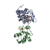
| Title | Crystal structure of the computationally designed NNOS-Syntrophin complex | ||||||
 Components Components |
| ||||||
 Keywords Keywords | MEMBRANE PROTEIN/OXIDOREDUCTASE / PDZ / Protein Binding / Dimerization / Mutation / Membrane / MEMBRANE PROTEIN-OXIDOREDUCTASE complex | ||||||
| Function / homology |  Function and homology information Function and homology informationregulation of vasoconstriction by circulating norepinephrine / dystrophin-associated glycoprotein complex / Formation of the dystrophin-glycoprotein complex (DGC) / dystroglycan binding / regulation of sodium ion transmembrane transport / ventricular cardiac muscle cell action potential / regulation of ventricular cardiac muscle cell membrane repolarization / Nitric oxide stimulates guanylate cyclase / negative regulation of hepatic stellate cell contraction / positive regulation of adenylate cyclase-activating adrenergic receptor signaling pathway ...regulation of vasoconstriction by circulating norepinephrine / dystrophin-associated glycoprotein complex / Formation of the dystrophin-glycoprotein complex (DGC) / dystroglycan binding / regulation of sodium ion transmembrane transport / ventricular cardiac muscle cell action potential / regulation of ventricular cardiac muscle cell membrane repolarization / Nitric oxide stimulates guanylate cyclase / negative regulation of hepatic stellate cell contraction / positive regulation of adenylate cyclase-activating adrenergic receptor signaling pathway / negative regulation of iron ion transmembrane transport / anchoring junction / response to vitamin B3 / ROS and RNS production in phagocytes / postsynaptic specialization, intracellular component / azurophil granule / Ion homeostasis / synaptic signaling by nitric oxide / negative regulation of vasoconstriction / response to nitric oxide / positive regulation of adenylate cyclase-activating G protein-coupled receptor signaling pathway / positive regulation of sodium ion transmembrane transport / response to vitamin E / peptidyl-cysteine S-nitrosylase activity / negative regulation of cytosolic calcium ion concentration / neuromuscular junction development / positive regulation of the force of heart contraction / cadmium ion binding / neuron projection terminus / negative regulation of calcium ion transport / negative regulation of potassium ion transport / nitric-oxide synthase binding / regulation of postsynaptic membrane potential / nitric-oxide synthase (NADPH) / sodium channel regulator activity / regulation of neurogenesis / : / nitric-oxide synthase activity / negative regulation of serotonin uptake / xenobiotic catabolic process / L-arginine catabolic process / NADPH binding / multicellular organismal response to stress / nitric oxide-cGMP-mediated signaling / postsynaptic density, intracellular component / regulation of sodium ion transport / nitric oxide metabolic process / striated muscle contraction / nitric oxide biosynthetic process / negative regulation of blood pressure / behavioral response to cocaine / photoreceptor inner segment / response to hormone / sarcoplasmic reticulum membrane / cellular response to epinephrine stimulus / establishment of localization in cell / T-tubule / secretory granule / regulation of heart rate / calyx of Held / response to activity / cell periphery / positive regulation of long-term synaptic potentiation / sarcoplasmic reticulum / response to nicotine / PDZ domain binding / neuromuscular junction / establishment of protein localization / female pregnancy / cellular response to mechanical stimulus / phosphoprotein binding / response to nutrient levels / sarcolemma / negative regulation of insulin secretion / caveola / response to estrogen / response to lead ion / cellular response to growth factor stimulus / response to peptide hormone / vasodilation / Z disc / calcium-dependent protein binding / NADP binding / FMN binding / flavin adenine dinucleotide binding / positive regulation of neuron apoptotic process / ATPase binding / response to heat / actin binding / scaffold protein binding / nuclear membrane / response to lipopolysaccharide / dendritic spine / cytoskeleton / RNA polymerase II-specific DNA-binding transcription factor binding / response to ethanol / negative regulation of neuron apoptotic process / transmembrane transporter binding / perikaryon / response to hypoxia Similarity search - Function | ||||||
| Biological species |  | ||||||
| Method |  X-RAY DIFFRACTION / SYNCHROTRON / MOLECULAR REPLACEMENT / Resolution: 2.29 Å X-RAY DIFFRACTION / SYNCHROTRON / MOLECULAR REPLACEMENT / Resolution: 2.29 Å | ||||||
 Authors Authors | Harwood, I.M. / Melero, C. / Ollikainen, N. / Kortemme, T. | ||||||
 Citation Citation | Journal: Proc.Natl.Acad.Sci.USA / Year: 2014 Title: Quantification of the transferability of a designed protein specificity switch reveals extensive epistasis in molecular recognition. Authors: Melero, C. / Ollikainen, N. / Harwood, I. / Karpiak, J. / Kortemme, T. | ||||||
| History |
|
- Structure visualization
| Structure viewer | Molecule: MolmilJmol/JSmol |
|---|
- Downloads & links
-Download
| PDBx/mmCIF format | 4hop.cif.gz | 136.3 KB | Display | PDBx/mmCIF format |
|---|---|---|---|---|
| PDB format | pdb4hop.ent.gz | 107.3 KB | Display | PDB format |
| PDBx/mmJSON format | 4hop.json.gz | Tree view | PDBx/mmJSON format | |
| Others |  Other downloads Other downloads |
-Validation report
| Arichive directory | https://data.pdbj.org/pub/pdb/validation_reports/ho/4hopftp://data.pdbj.org/pub/pdb/validation_reports/ho/4hop | HTTPS FTP |
|---|
-Related structure data
| Related structure data |  1qavS S: Starting model for refinement |
|---|---|
| Similar structure data |


-Links
 PDBj
PDBj


- Assembly
| Deposited unit | 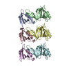
| ||||||||
|---|---|---|---|---|---|---|---|---|---|
| 1 | 
| ||||||||
| 2 | 
| ||||||||
| 3 | 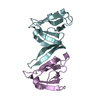
| ||||||||
| Unit cell |
|
-Components
| #1: Protein | Mass: 9389.835 Da / Num. of mol.: 3 / Fragment: PDZ DOMAIN (RESIDUES 77-162) / Mutation: H142F Source method: isolated from a genetically manipulated source Source: (gene. exp.)  #2: Protein | Mass: 13289.354 Da / Num. of mol.: 3 / Fragment: PDZ DOMAIN (RESIDUES 4-126) / Mutation: T109M Source method: isolated from a genetically manipulated source Source: (gene. exp.) #3: Water | ChemComp-HOH / |  Mass: 18.015 Da / Num. of mol.: 501 / Source method: isolated from a natural source / Formula: H2O Mass: 18.015 Da / Num. of mol.: 501 / Source method: isolated from a natural source / Formula: H2O |
|---|
-Experimental details
-Experiment
| Experiment | Method: X-RAY DIFFRACTION / Number of used crystals: 1 |
|---|
- Sample preparation
| Crystal | Density Matthews: 2.59 Å3/Da / Density % sol: 52.58 % |
|---|---|
| Crystal grow | Temperature: 293 K / Method: vapor diffusion, hanging drop / pH: 5.25 Details: 0.1M MES, 0.2M LICL, 21.5% PEG 6000. SYNTROPHIN AT 3.6 MG/ML FINAL. NNOS AT 2.6 MG/ML FINAL. 1:1 MIX OF PROTEIN SOLUTION TO PRECIPITANT, pH 5.25, VAPOR DIFFUSION, HANGING DROP, temperature 293K |
-Data collection
| Diffraction | Mean temperature: 77.2 K |
|---|---|
| Diffraction source | Source: SYNCHROTRON / Site: ALS  / Beamline: 8.3.1 / Wavelength: 1.11587 / Beamline: 8.3.1 / Wavelength: 1.11587 |
| Detector | Type: ADSC QUANTUM 315 / Detector: CCD / Date: Jun 29, 2007 Details: MIRROR1: PLANE PARABOLA PT AND RH-COATED INVAR STEEL, MIRROR2: TOROID (2:1 DEMAGNIFICATION) PT AND RH- COATED SI |
| Radiation | Monochromator: KOHZU DOUBLE FLAT SI(111) CRYSTAL / Protocol: SINGLE WAVELENGTH / Monochromatic (M) / Laue (L): M / Scattering type: x-ray |
| Radiation wavelength | Wavelength: 1.11587 Å / Relative weight: 1 |
| Reflection | Resolution: 2.29→48 Å / Num. obs: 31327 / % possible obs: 100 % / Observed criterion σ(I): -3 / Redundancy: 9.1 % / Rmerge(I) obs: 0.068 / Rsym value: 0.068 / Net I/σ(I): 30.532 |
| Reflection shell | Resolution: 2.29→2.37 Å / Redundancy: 6 % / Rmerge(I) obs: 0.171 / Mean I/σ(I) obs: 12.043 / Rsym value: 0.171 / % possible all: 99.8 |
- Processing
| Software |
| ||||||||||||||||||||||||||||||||||||||||||||||||||||||||||||||||||||||||||||||||||||||||||||||||||||||||||||||||||||||||||||||||||||||||||||||||||||||||||||||||||||||||||
|---|---|---|---|---|---|---|---|---|---|---|---|---|---|---|---|---|---|---|---|---|---|---|---|---|---|---|---|---|---|---|---|---|---|---|---|---|---|---|---|---|---|---|---|---|---|---|---|---|---|---|---|---|---|---|---|---|---|---|---|---|---|---|---|---|---|---|---|---|---|---|---|---|---|---|---|---|---|---|---|---|---|---|---|---|---|---|---|---|---|---|---|---|---|---|---|---|---|---|---|---|---|---|---|---|---|---|---|---|---|---|---|---|---|---|---|---|---|---|---|---|---|---|---|---|---|---|---|---|---|---|---|---|---|---|---|---|---|---|---|---|---|---|---|---|---|---|---|---|---|---|---|---|---|---|---|---|---|---|---|---|---|---|---|---|---|---|---|---|---|---|---|
| Refinement | Method to determine structure: MOLECULAR REPLACEMENT Starting model: PDB ENTRY 1QAV Resolution: 2.29→47.57 Å / Cor.coef. Fo:Fc: 0.93 / Cor.coef. Fo:Fc free: 0.9 / SU B: 7.709 / SU ML: 0.191 / Cross valid method: THROUGHOUT / ESU R: 0.424 / ESU R Free: 0.273 / Stereochemistry target values: MAXIMUM LIKELIHOOD / Details: HYDROGENS HAVE BEEN ADDED IN THE RIDING POSITIONS
| ||||||||||||||||||||||||||||||||||||||||||||||||||||||||||||||||||||||||||||||||||||||||||||||||||||||||||||||||||||||||||||||||||||||||||||||||||||||||||||||||||||||||||
| Solvent computation | Ion probe radii: 0.8 Å / Shrinkage radii: 0.8 Å / VDW probe radii: 1.2 Å / Solvent model: MASK | ||||||||||||||||||||||||||||||||||||||||||||||||||||||||||||||||||||||||||||||||||||||||||||||||||||||||||||||||||||||||||||||||||||||||||||||||||||||||||||||||||||||||||
| Displacement parameters | Biso mean: 15.81 Å2
| ||||||||||||||||||||||||||||||||||||||||||||||||||||||||||||||||||||||||||||||||||||||||||||||||||||||||||||||||||||||||||||||||||||||||||||||||||||||||||||||||||||||||||
| Refinement step | Cycle: LAST / Resolution: 2.29→47.57 Å
| ||||||||||||||||||||||||||||||||||||||||||||||||||||||||||||||||||||||||||||||||||||||||||||||||||||||||||||||||||||||||||||||||||||||||||||||||||||||||||||||||||||||||||
| Refine LS restraints |
| ||||||||||||||||||||||||||||||||||||||||||||||||||||||||||||||||||||||||||||||||||||||||||||||||||||||||||||||||||||||||||||||||||||||||||||||||||||||||||||||||||||||||||
| LS refinement shell | Resolution: 2.29→2.35 Å / Total num. of bins used: 20
|
