 ムービー
ムービー コントローラー


+ データを開く

- 基本情報
基本情報
| 登録情報 | データベース: PDB / ID: 2jqr | ||||||
|---|---|---|---|---|---|---|---|
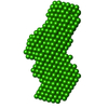
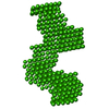
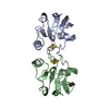
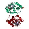
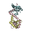
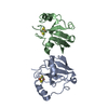
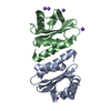
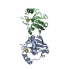
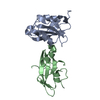
| タイトル | Solution model of crosslinked complex of cytochrome c and adrenodoxin | ||||||
 要素 要素 |
| ||||||
 キーワード キーワード | ELECTRON TRANSPORT / Cytochrome c / Adrenodoxin / Crosslinked complex / 2Fe2S Ferredoxin / Pseudocontact shift / Paramagnetic relaxation enhancement / encounter complex | ||||||
| 機能・相同性 |  機能・相同性情報 機能・相同性情報Mitochondrial iron-sulfur cluster biogenesis / Pregnenolone biosynthesis / Electron transport from NADPH to Ferredoxin / Endogenous sterols / Protein lipoylation / hormone biosynthetic process / Release of apoptotic factors from the mitochondria / Pyroptosis / P450-containing electron transport chain / Detoxification of Reactive Oxygen Species ...Mitochondrial iron-sulfur cluster biogenesis / Pregnenolone biosynthesis / Electron transport from NADPH to Ferredoxin / Endogenous sterols / Protein lipoylation / hormone biosynthetic process / Release of apoptotic factors from the mitochondria / Pyroptosis / P450-containing electron transport chain / Detoxification of Reactive Oxygen Species / Respiratory electron transport / steroid biosynthetic process / cardiolipin binding / mitochondrial electron transport, cytochrome c to oxygen / mitochondrial electron transport, ubiquinol to cytochrome c / cholesterol metabolic process / cellular response to cAMP / cellular response to forskolin / respiratory electron transport chain / electron transport chain / mitochondrial intermembrane space / 2 iron, 2 sulfur cluster binding / electron transfer activity / mitochondrial matrix / heme binding / protein homodimerization activity / mitochondrion / metal ion binding 類似検索 - 分子機能 | ||||||
| 生物種 |   | ||||||
| 手法 | 溶液NMR / molecular dynamics | ||||||
 データ登録者 データ登録者 | Xu, X. / Reinle, W. / Hannemann, F. / Konarev, P.V. / Svergun, D.I. / Bernhardt, R. / Ubbink, M. | ||||||
 引用 引用 | ジャーナル: J Am Chem Soc / 年: 2008 タイトル: Dynamics in a pure encounter complex of two proteins studied by solution scattering and paramagnetic NMR spectroscopy. 著者: Xingfu Xu / Wolfgang Reinle / Frank Hannemann / Peter V Konarev / Dmitri I Svergun / Rita Bernhardt / Marcellus Ubbink /  要旨: In the general view of protein-complex formation, a transient and dynamic encounter complex proceeds to form a more stable, well-defined, and active form. In weak protein complexes, however, the ...In the general view of protein-complex formation, a transient and dynamic encounter complex proceeds to form a more stable, well-defined, and active form. In weak protein complexes, however, the encounter state can represent a significant population of the complex. The redox proteins adrenodoxin (Adx) and cytochrome c (C c) associate to form such a weak and short-lived complex, which is nevertheless active in electron transfer. To study the conformational freedom within the protein complex, the native complex has been compared to a cross-linked counterpart by using solution scattering and NMR spectroscopy. Oligomerization behavior of the native complex in solution revealed by small-angle X-ray scattering indicates a stochastic nature of complex formation. For the cross-linked complex, interprotein paramagnetic effects are observed, whereas for the native complex, extensive averaging occurs, consistent with multiple orientations of the proteins within the complex. Simulations show that C c samples about half of the surface area of adrenodoxin. It is concluded that the complex of Adx/C c is entirely dynamic and can be considered as a pure encounter complex. #1: ジャーナル: J.Mol.Biol. / 年: 1990タイトル: High-resolution refinement of yeast iso-1-cytochrome C and comparisons with other eukaryotic cytochromes C 著者: Louie, G.V. / Brayer, G.D. #2: ジャーナル: Structure / 年: 1998タイトル: New aspects of electron transfer revealed by the crystal structure of a truncated bovine adrenodoxin, Adx(4-108) 著者: Muller, A. / Muller, J.J. / Muller, Y.A. / Uhlmann, H. / Bernhardt, R. / Heinemann, U. | ||||||
| 履歴 |
|
- 構造の表示
構造の表示
| 構造ビューア | 分子: MolmilJmol/JSmol |
|---|
- ダウンロードとリンク
-ダウンロード
| PDBx/mmCIF形式 | 2jqr.cif.gz | 659.5 KB | 表示 | PDBx/mmCIF形式 |
|---|---|---|---|---|
| PDB形式 | pdb2jqr.ent.gz | 549.7 KB | 表示 | PDB形式 |
| PDBx/mmJSON形式 | 2jqr.json.gz | ツリー表示 | PDBx/mmJSON形式 | |
| その他 |  その他のダウンロード その他のダウンロード |
-検証レポート
| アーカイブディレクトリ | https://data.pdbj.org/pub/pdb/validation_reports/jq/2jqrftp://data.pdbj.org/pub/pdb/validation_reports/jq/2jqr | HTTPS FTP |
|---|
-関連構造データ
| 関連構造データ | C: 同じ文献を引用 ( |
|---|---|
| 類似構造データ | |
| その他のデータベース |


-リンク
 PDBj
PDBj


- 集合体
集合体
| 登録構造単位 | 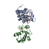
| |||||||||
|---|---|---|---|---|---|---|---|---|---|---|
| 1 |
| |||||||||
| NMR アンサンブル |
|
-要素
| #1: タンパク質 | 分子量: 12075.808 Da / 分子数: 1 / 変異: V28C, C102T / 由来タイプ: 組換発現 由来: (組換発現) 遺伝子: CYC1 / 生物種 (発現宿主): Escherichia coli / 発現宿主:  |
|---|---|
| #2: タンパク質 | 分子量: 11635.125 Da / 分子数: 1 / Fragment: 2Fe-2S ferredoxin-type domain, residues 62-166 / 変異: L80C, C95S / 由来タイプ: 組換発現 / 由来: (組換発現) |
| #3: 化合物 | ChemComp-HEC /   分子量: 618.503 Da / 分子数: 1 / 由来タイプ: 合成 / 式: C34H34FeN4O4 分子量: 618.503 Da / 分子数: 1 / 由来タイプ: 合成 / 式: C34H34FeN4O4 |
| #4: 化合物 | ChemComp-FES /   分子量: 175.820 Da / 分子数: 1 / 由来タイプ: 合成 / 式: Fe2S2 分子量: 175.820 Da / 分子数: 1 / 由来タイプ: 合成 / 式: Fe2S2 |
| Has protein modification | Y |
-実験情報
-実験
| 実験 | 手法: 溶液NMR | ||||||||||||||||||||||||||||||||||||
|---|---|---|---|---|---|---|---|---|---|---|---|---|---|---|---|---|---|---|---|---|---|---|---|---|---|---|---|---|---|---|---|---|---|---|---|---|---|
| NMR実験 |
|
 HSQC
HSQC- 試料調製
試料調製
| 詳細 |
| ||||||||||||||||||||||||||||||||||||||||||||||||||||
|---|---|---|---|---|---|---|---|---|---|---|---|---|---|---|---|---|---|---|---|---|---|---|---|---|---|---|---|---|---|---|---|---|---|---|---|---|---|---|---|---|---|---|---|---|---|---|---|---|---|---|---|---|---|
| 試料 |
| ||||||||||||||||||||||||||||||||||||||||||||||||||||
| 試料状態 |
|
-NMR測定
| NMRスペクトロメーター | タイプ: Bruker DMX / 製造業者: Bruker / モデル: DMX / 磁場強度: 600 MHz |
|---|
- 解析
解析
| NMR software |
| ||||||||||||
|---|---|---|---|---|---|---|---|---|---|---|---|---|---|
| 精密化 | 手法: molecular dynamics / ソフトェア番号: 1 詳細: All structure models are from rigid body modeling. The coordinate of Cytochrome C (Chain A) is from PDB entry 1YCC. All complex structure models are superimposed with Chain A. | ||||||||||||
| 代表構造 | 選択基準: lowest energy | ||||||||||||
| NMRアンサンブル | コンフォーマー選択の基準: structures with the lowest energy 計算したコンフォーマーの数: 10 / 登録したコンフォーマーの数: 10 |
