Monochromator: SAGITALLY FOCUSED SI(111) / Protocol: SINGLE WAVELENGTH / Monochromatic (M) / Laue (L): M / Scattering type: x-ray
Radiation wavelength
Wavelength: 0.97949 Å / Relative weight: 1
Reflection
Redundancy: 16.3 % / Av σ(I) over netI: 10.79 / Number: 343790 / Rmerge(I) obs: 0.188 / Χ2: 1.29 / D res high: 3.3 Å / D res low: 40 Å / Num. obs: 21100 / % possible obs: 100
Diffraction reflection shell
Highest resolution (Å)
Lowest resolution (Å)
% possible obs (%)
ID
Rmerge(I) obs
Chi squared
Redundancy
8.93
40
100
1
0.064
3.102
14
7.1
8.93
100
1
0.082
1.863
15
6.2
7.1
100
1
0.122
1.464
15.7
5.64
6.2
100
1
0.132
1.219
16
5.24
5.64
100
1
0.138
1.219
16.3
4.93
5.24
100
1
0.123
1.165
16.3
4.68
4.93
100
1
0.128
1.201
16.3
4.48
4.68
100
1
0.141
1.163
16.6
4.31
4.48
100
1
0.154
1.169
16.5
4.16
4.31
100
1
0.181
1.164
16.6
4.03
4.16
100
1
0.214
1.13
16.7
3.91
4.03
100
1
0.241
1.127
16.7
3.81
3.91
100
1
0.265
1.15
16.7
3.72
3.81
100
1
0.296
1.16
16.9
3.63
3.72
100
1
0.352
1.107
16.7
3.55
3.63
100
1
0.389
1.079
16.9
3.48
3.55
100
1
0.424
1.083
16.8
3.42
3.48
100
1
0.488
1.057
16.8
3.36
3.42
100
1
0.599
1.077
16.4
3.3
3.36
100
1
0.648
1.156
16.4
Reflection
Resolution: 2.84→50 Å / Num. obs: 32326 / % possible obs: 99.8 % / Observed criterion σ(I): 3 / Redundancy: 5.9 % / Rmerge(I) obs: 0.133 / Net I/σ(I): 5.6
Reflection shell
Resolution: 2.84→2.89 Å / Redundancy: 5.9 % / % possible all: 100
-
Phasing
Phasing
Method: SAD
-
Processing
Software
Name
Version
Classification
NB
DENZO
datareduction
SCALEPACK
datascaling
SHELX
phasing
DM
phasing
PHENIX
1.4_175
refinement
PDB_EXTRACT
3.1
dataextraction
HKL-2000
datareduction
HKL-2000
datascaling
SHARP
phasing
Refinement
Method to determine structure: SAD / Resolution: 3→39.2 Å / Occupancy max: 1 / Occupancy min: 1 / SU ML: 0.34 / Isotropic thermal model: TLS / σ(F): 0.2 / Phase error: 21.91 / Stereochemistry target values: ML
Rfactor
Num. reflection
% reflection
Rfree
0.242
2475
9.52 %
Rwork
0.17
-
-
obs
0.176
25991
94.6 %
Solvent computation
Shrinkage radii: 0.9 Å / VDW probe radii: 1.11 Å / Solvent model: FLAT BULK SOLVENT MODEL / Bsol: 28.21 Å2 / ksol: 0.33 e/Å3
Displacement parameters
Biso mean: 59.18 Å2
Baniso -1
Baniso -2
Baniso -3
1-
5.837 Å2
0 Å2
-0 Å2
2-
-
5.837 Å2
-0 Å2
3-
-
-
-11.673 Å2
Refinement step
Cycle: LAST / Resolution: 3→39.2 Å
Protein
Nucleic acid
Ligand
Solvent
Total
Num. atoms
6590
0
66
52
6708
Refine LS restraints
Refine-ID
Type
Dev ideal
Number
X-RAY DIFFRACTION
f_bond_d
0.009
6829
X-RAY DIFFRACTION
f_angle_d
1.215
9318
X-RAY DIFFRACTION
f_dihedral_angle_d
18.813
2438
X-RAY DIFFRACTION
f_chiral_restr
0.076
1062
X-RAY DIFFRACTION
f_plane_restr
0.005
1215
LS refinement shell
Resolution (Å)
Rfactor Rfree
Num. reflection Rfree
Rfactor Rwork
Num. reflection Rwork
Refine-ID
% reflection obs (%)
3-3.0578
0.3363
127
0.2398
1102
X-RAY DIFFRACTION
82
3.0578-3.1202
0.3506
120
0.2435
1187
X-RAY DIFFRACTION
88
3.1202-3.188
0.3164
139
0.2074
1151
X-RAY DIFFRACTION
87
3.188-3.2621
0.2948
123
0.1933
1249
X-RAY DIFFRACTION
91
3.2621-3.3436
0.2302
128
0.177
1248
X-RAY DIFFRACTION
92
3.3436-3.434
0.2912
126
0.1792
1270
X-RAY DIFFRACTION
94
3.434-3.535
0.2648
130
0.1755
1297
X-RAY DIFFRACTION
94
3.535-3.649
0.2365
139
0.1656
1289
X-RAY DIFFRACTION
96
3.649-3.7793
0.237
142
0.1605
1302
X-RAY DIFFRACTION
97
3.7793-3.9305
0.2114
135
0.147
1322
X-RAY DIFFRACTION
96
3.9305-4.1092
0.2749
142
0.1491
1320
X-RAY DIFFRACTION
97
4.1092-4.3256
0.207
139
0.1335
1330
X-RAY DIFFRACTION
97
4.3256-4.5962
0.2107
141
0.1323
1373
X-RAY DIFFRACTION
98
4.5962-4.9505
0.1952
144
0.1252
1365
X-RAY DIFFRACTION
98
4.9505-5.4475
0.2184
146
0.1378
1363
X-RAY DIFFRACTION
98
5.4475-6.233
0.2073
149
0.1477
1394
X-RAY DIFFRACTION
98
6.233-7.8426
0.2256
144
0.1515
1424
X-RAY DIFFRACTION
98
7.8426-39.1985
0.1917
161
0.1772
1530
X-RAY DIFFRACTION
99
Refinement TLS params.
Method: refined / Refine-ID: X-RAY DIFFRACTION
ID
L11 (°2)
L12 (°2)
L13 (°2)
L22 (°2)
L23 (°2)
L33 (°2)
S11 (Å °)
S12 (Å °)
S13 (Å °)
S21 (Å °)
S22 (Å °)
S23 (Å °)
S31 (Å °)
S32 (Å °)
S33 (Å °)
T11 (Å2)
T12 (Å2)
T13 (Å2)
T22 (Å2)
T23 (Å2)
T33 (Å2)
Origin x (Å)
Origin y (Å)
Origin z (Å)
1
0.6428
-0.2938
-0.7617
1.0263
1.1885
3.4038
0.0349
-0.0057
0.1703
-0.1346
0.2166
-0.2971
-0.7769
0.4335
-0
0.3971
-0.0104
-0.0079
0.2577
-0.0538
0.3304
28.9326
62.9562
139.5017
2
1.1336
0.724
-0.167
2.6713
0.7945
1.9075
-0.048
-0.0356
0.0945
-0.2641
0.1929
-0.0635
-0.2167
0.1884
-0.0001
0.0879
0.0125
0.0135
0.2388
-0.0599
0.2533
31.6204
38.344
114.5897
3
3.2505
0.4657
0.5087
3.8159
0.6215
1.3625
0.0932
0.132
-0.4528
-0.1675
-0.0991
0.0631
0.2207
0.0131
0.0005
0.0821
0.0661
0.0202
0.2454
-0.07
0.2324
23.2648
10.0059
112.2203
4
0.3058
0.3214
0.0454
0.3697
-0.0692
0.3235
-0.1826
-0.0598
-0.7664
0.677
-0.1104
0.4306
0.3906
-0.3443
-0.0012
0.4222
0.0513
0.1001
0.5731
-0.0729
0.4928
9.7812
58.0508
155.9645
5
1.7977
-0.6741
-0.4096
1.6849
0.7457
1.8627
-0.0822
0.0891
-0.052
-0.2628
0.1718
-0.0908
-0.4715
0.0765
-0.0002
0.3868
0.1255
0.0278
0.1814
-0.0542
0.1243
21.6799
63.4953
150.7866
Refinement TLS group
ID
Refine-ID
Refine TLS-ID
Selection details
1
X-RAY DIFFRACTION
1
(CHAINAANDRESID49:303)
2
X-RAY DIFFRACTION
2
(CHAINAANDRESID304:614)
3
X-RAY DIFFRACTION
3
(CHAINAANDRESID615:797)
4
X-RAY DIFFRACTION
4
(CHAINBANDRESID24:51)
5
X-RAY DIFFRACTION
5
(CHAINBANDRESID52:165)
+
About Yorodumi
-
News
-
Feb 9, 2022. New format data for meta-information of EMDB entries
New format data for meta-information of EMDB entries
Version 3 of the EMDB header file is now the official format.
The previous official version 1.9 will be removed from the archive.
In the structure databanks used in Yorodumi, some data are registered as the other names, "COVID-19 virus" and "2019-nCoV". Here are the details of the virus and the list of structure data.
Jan 31, 2019. EMDB accession codes are about to change! (news from PDBe EMDB page)
EMDB accession codes are about to change! (news from PDBe EMDB page)
The allocation of 4 digits for EMDB accession codes will soon come to an end. Whilst these codes will remain in use, new EMDB accession codes will include an additional digit and will expand incrementally as the available range of codes is exhausted. The current 4-digit format prefixed with “EMD-” (i.e. EMD-XXXX) will advance to a 5-digit format (i.e. EMD-XXXXX), and so on. It is currently estimated that the 4-digit codes will be depleted around Spring 2019, at which point the 5-digit format will come into force.
The EM Navigator/Yorodumi systems omit the EMD- prefix.
Related info.:Q: What is EMD? / ID/Accession-code notation in Yorodumi/EM Navigator
Yorodumi is a browser for structure data from EMDB, PDB, SASBDB, etc.
This page is also the successor to EM Navigator detail page, and also detail information page/front-end page for Omokage search.
The word "yorodu" (or yorozu) is an old Japanese word meaning "ten thousand". "mi" (miru) is to see.
Related info.:EMDB / PDB / SASBDB / Comparison of 3 databanks / Yorodumi Search / Aug 31, 2016. New EM Navigator & Yorodumi / Yorodumi Papers / Jmol/JSmol / Function and homology information / Changes in new EM Navigator and Yorodumi
 Movie
Movie Controller

 Yorodumi
 Open data
 Basic information
 Components
 Keywords
 Function and homology information
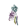
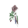
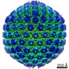 Human herpesvirus 2
 X-RAY DIFFRACTION /
  Authors
 Citation
 Structure visualization
 Downloads & links
 Other downloads
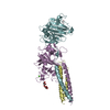


 PDBj
PDBj Assembly

 Spodoptera frugiperda (fall armyworm) / Strain (production host): Sf9 / References: UniProt: P89445

 Type: D-saccharide, beta linking / Mass: 221.208 Da / Num. of mol.: 2 / Source method: isolated from a natural source / Formula: C8H15NO6
 Type: D-saccharide / Mass: 152.146 Da / Num. of mol.: 1
 Sample preparation
 / Beamline: 24-ID-C / Wavelength: 0.97949
  Processing