 Movie
Movie Controller

[English] 日本語
 Yorodumi
- PDB-3e1i: Crystal Structure of BbetaD432A Variant Fibrinogen Fragment D wit... -
+ Open data

- Basic information
| Entry | Database: PDB / ID: 3e1i | ||||||
|---|---|---|---|---|---|---|---|
| Title | Crystal Structure of BbetaD432A Variant Fibrinogen Fragment D with the Peptide Ligand Gly-His-Arg-Pro-amide | ||||||
 Components Components |
| ||||||
 Keywords Keywords | BLOOD CLOTTING / Blood Coagulation / Disease mutation / Glycoprotein / Phosphoprotein / Secreted / Pyrrolidone carboxylic acid / Sulfation | ||||||
| Function / homology |  Function and homology information Function and homology informationblood coagulation, common pathway / induction of bacterial agglutination / fibrinogen complex / Regulation of TLR by endogenous ligand / platelet alpha granule / cellular response to leptin stimulus / MyD88 deficiency (TLR2/4) / positive regulation of heterotypic cell-cell adhesion / IRAK4 deficiency (TLR2/4) / plasminogen activation ...blood coagulation, common pathway / induction of bacterial agglutination / fibrinogen complex / Regulation of TLR by endogenous ligand / platelet alpha granule / cellular response to leptin stimulus / MyD88 deficiency (TLR2/4) / positive regulation of heterotypic cell-cell adhesion / IRAK4 deficiency (TLR2/4) / plasminogen activation / MyD88:MAL(TIRAP) cascade initiated on plasma membrane / extracellular matrix structural constituent / p130Cas linkage to MAPK signaling for integrins / positive regulation of peptide hormone secretion / GRB2:SOS provides linkage to MAPK signaling for Integrins / positive regulation of exocytosis / blood coagulation, fibrin clot formation / protein polymerization / protein secretion / positive regulation of vasoconstriction / cellular response to interleukin-1 / Integrin cell surface interactions / : / negative regulation of endothelial cell apoptotic process / negative regulation of extrinsic apoptotic signaling pathway via death domain receptors / fibrinolysis / Integrin signaling / positive regulation of substrate adhesion-dependent cell spreading / cell adhesion molecule binding / platelet alpha granule lumen / cell-matrix adhesion / positive regulation of protein secretion / Post-translational protein phosphorylation / Signaling by high-kinase activity BRAF mutants / MAP2K and MAPK activation / response to calcium ion / platelet aggregation / Regulation of Insulin-like Growth Factor (IGF) transport and uptake by Insulin-like Growth Factor Binding Proteins (IGFBPs) / Signaling by RAF1 mutants / Signaling by moderate kinase activity BRAF mutants / Paradoxical activation of RAF signaling by kinase inactive BRAF / Signaling downstream of RAS mutants / Signaling by BRAF and RAF1 fusions / Platelet degranulation / extracellular vesicle / protein-folding chaperone binding / extracellular matrix / ER-Phagosome pathway / protein-containing complex assembly / blood microparticle / cell cortex / adaptive immune response / protein-macromolecule adaptor activity / positive regulation of ERK1 and ERK2 cascade / endoplasmic reticulum lumen / Amyloid fiber formation / signaling receptor binding / external side of plasma membrane / innate immune response / synapse / structural molecule activity / cell surface / endoplasmic reticulum / : / extracellular exosome / extracellular region / metal ion binding / plasma membrane Similarity search - Function | ||||||
| Biological species |  Homo sapiens (human) Homo sapiens (human)synthetic construct (others) | ||||||
| Method |  X-RAY DIFFRACTION / SYNCHROTRON / MOLECULAR REPLACEMENT / Resolution: 2.3 Å X-RAY DIFFRACTION / SYNCHROTRON / MOLECULAR REPLACEMENT / Resolution: 2.3 Å | ||||||
 Authors Authors | Bowley, S.R. / Lord, S.T. | ||||||
 Citation Citation | Journal: Blood / Year: 2009 Title: Fibrinogen variant BbetaD432A has normal polymerization but does not bind knob "B". Authors: Bowley, S.R. / Lord, S.T. | ||||||
| History |
|
- Structure visualization
| Structure viewer | Molecule: MolmilJmol/JSmol |
|---|
- Downloads & links
-Download
| PDBx/mmCIF format | 3e1i.cif.gz | 276 KB | Display | PDBx/mmCIF format |
|---|---|---|---|---|
| PDB format | pdb3e1i.ent.gz | 219.4 KB | Display | PDB format |
| PDBx/mmJSON format | 3e1i.json.gz | Tree view | PDBx/mmJSON format | |
| Others |  Other downloads Other downloads |
-Validation report
| Arichive directory | https://data.pdbj.org/pub/pdb/validation_reports/e1/3e1iftp://data.pdbj.org/pub/pdb/validation_reports/e1/3e1i | HTTPS FTP |
|---|
-Related structure data
| Related structure data |  1lt9S S: Starting model for refinement |
|---|---|
| Similar structure data |


-Links
 PDBj
PDBj


- Assembly
| Deposited unit | 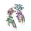
| ||||||||
|---|---|---|---|---|---|---|---|---|---|
| 1 | 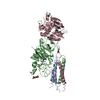
| ||||||||
| 2 | 
| ||||||||
| Unit cell |
|
-Components
-Protein , 3 types, 6 molecules ADBECF
| #1: Protein | Mass: 10244.963 Da / Num. of mol.: 2 Source method: isolated from a genetically manipulated source Source: (gene. exp.) Homo sapiens (human) / Gene: FGA / Plasmid: pMLP / Cell line (production host): CHO / Production host:   Cricetulus griseus (Chinese hamster) / References: UniProt: P02671 Cricetulus griseus (Chinese hamster) / References: UniProt: P02671#2: Protein | Mass: 37647.984 Da / Num. of mol.: 2 / Mutation: D432A Source method: isolated from a genetically manipulated source Source: (gene. exp.) Homo sapiens (human) / Gene: FGB / Plasmid: pMLP / Cell line (production host): CHO / Production host: Cricetulus griseus (Chinese hamster) / References: UniProt: P02675#3: Protein | Mass: 36223.281 Da / Num. of mol.: 2 Source method: isolated from a genetically manipulated source Source: (gene. exp.) Homo sapiens (human) / Gene: FGG, PRO2061 / Plasmid: pMLP / Cell line (production host): CHO / Production host: Cricetulus griseus (Chinese hamster) / References: UniProt: P02679 |
|---|
-Protein/peptide / Sugars , 2 types, 4 molecules GH
| #4: Protein/peptide | Mass: 465.530 Da / Num. of mol.: 2 / Source method: obtained synthetically / Source: (synth.) synthetic construct (others) #5: Sugar |  Type: D-saccharide, beta linking / Mass: 221.208 Da / Num. of mol.: 2 Type: D-saccharide, beta linking / Mass: 221.208 Da / Num. of mol.: 2Source method: isolated from a genetically manipulated source Formula: C8H15NO6 |
|---|
-Non-polymers , 2 types, 267 molecules 

| #6: Chemical | ChemComp-CA /  Mass: 40.078 Da / Num. of mol.: 7 / Source method: obtained synthetically / Formula: Ca Mass: 40.078 Da / Num. of mol.: 7 / Source method: obtained synthetically / Formula: Ca#7: Water | ChemComp-HOH / | Mass: 18.015 Da / Num. of mol.: 260 / Source method: isolated from a natural source / Formula: H2O |
|---|
-Details
| Has protein modification | Y |
|---|
-Experimental details
-Experiment
| Experiment | Method: X-RAY DIFFRACTION / Number of used crystals: 1 |
|---|
- Sample preparation
| Crystal | Density Matthews: 2.7 Å3/Da / Density % sol: 54.48 % |
|---|---|
| Crystal grow | Temperature: 277 K / Method: vapor diffusion, sitting drop / pH: 8.5 Details: 50 mM Tris, 12.5 mM calcium chloride, 12% PEG 3350, 2 mM sodium azide, pH 8.5, VAPOR DIFFUSION, SITTING DROP, temperature 277K |
-Data collection
| Diffraction | Mean temperature: 100 K |
|---|---|
| Diffraction source | Source: SYNCHROTRON / Site: APS  / Beamline: 22-ID / Wavelength: 0.97625 Å / Beamline: 22-ID / Wavelength: 0.97625 Å |
| Detector | Type: MAR CCD 165 mm / Detector: CCD / Date: Aug 18, 2005 |
| Radiation | Protocol: SINGLE WAVELENGTH / Monochromatic (M) / Laue (L): M / Scattering type: x-ray |
| Radiation wavelength | Wavelength: 0.97625 Å / Relative weight: 1 |
| Reflection | Resolution: 2.15→50 Å / Num. obs: 82662 / % possible obs: 83 % / Observed criterion σ(F): 0 / Observed criterion σ(I): 0 / Redundancy: 6.7 % / Net I/σ(I): 29.5 |
| Reflection shell | Resolution: 2.15→2.28 Å / Redundancy: 5.9 % / Mean I/σ(I) obs: 3.4 / Rsym value: 0.284 / % possible all: 80.5 |
- Processing
| Software |
| |||||||||||||||||||||||||||||||||||||||||||||||||||||||||||||||||||||||||||||||||||||||||||||||||||||||||||||||||||||||||||||||||||||||||||||||||||||||||||||||||||||||||||||||
|---|---|---|---|---|---|---|---|---|---|---|---|---|---|---|---|---|---|---|---|---|---|---|---|---|---|---|---|---|---|---|---|---|---|---|---|---|---|---|---|---|---|---|---|---|---|---|---|---|---|---|---|---|---|---|---|---|---|---|---|---|---|---|---|---|---|---|---|---|---|---|---|---|---|---|---|---|---|---|---|---|---|---|---|---|---|---|---|---|---|---|---|---|---|---|---|---|---|---|---|---|---|---|---|---|---|---|---|---|---|---|---|---|---|---|---|---|---|---|---|---|---|---|---|---|---|---|---|---|---|---|---|---|---|---|---|---|---|---|---|---|---|---|---|---|---|---|---|---|---|---|---|---|---|---|---|---|---|---|---|---|---|---|---|---|---|---|---|---|---|---|---|---|---|---|---|---|
| Refinement | Method to determine structure: MOLECULAR REPLACEMENT Starting model: PDB ENTRY 1LT9 Resolution: 2.3→49.21 Å / Cor.coef. Fo:Fc: 0.936 / Cor.coef. Fo:Fc free: 0.918 / SU B: 11.139 / SU ML: 0.142 / TLS residual ADP flag: LIKELY RESIDUAL / Isotropic thermal model: restrained / Cross valid method: THROUGHOUT / ESU R: 0.275 / ESU R Free: 0.209 / Stereochemistry target values: MAXIMUM LIKELIHOOD / Details: HYDROGENS HAVE BEEN ADDED IN THE RIDING POSITIONS
| |||||||||||||||||||||||||||||||||||||||||||||||||||||||||||||||||||||||||||||||||||||||||||||||||||||||||||||||||||||||||||||||||||||||||||||||||||||||||||||||||||||||||||||||
| Solvent computation | Ion probe radii: 0.8 Å / Shrinkage radii: 0.8 Å / VDW probe radii: 1.2 Å / Solvent model: MASK | |||||||||||||||||||||||||||||||||||||||||||||||||||||||||||||||||||||||||||||||||||||||||||||||||||||||||||||||||||||||||||||||||||||||||||||||||||||||||||||||||||||||||||||||
| Displacement parameters | Biso mean: 33.558 Å2
| |||||||||||||||||||||||||||||||||||||||||||||||||||||||||||||||||||||||||||||||||||||||||||||||||||||||||||||||||||||||||||||||||||||||||||||||||||||||||||||||||||||||||||||||
| Refinement step | Cycle: LAST / Resolution: 2.3→49.21 Å
| |||||||||||||||||||||||||||||||||||||||||||||||||||||||||||||||||||||||||||||||||||||||||||||||||||||||||||||||||||||||||||||||||||||||||||||||||||||||||||||||||||||||||||||||
| Refine LS restraints |
| |||||||||||||||||||||||||||||||||||||||||||||||||||||||||||||||||||||||||||||||||||||||||||||||||||||||||||||||||||||||||||||||||||||||||||||||||||||||||||||||||||||||||||||||
| LS refinement shell | Resolution: 2.3→2.36 Å / Total num. of bins used: 20
| |||||||||||||||||||||||||||||||||||||||||||||||||||||||||||||||||||||||||||||||||||||||||||||||||||||||||||||||||||||||||||||||||||||||||||||||||||||||||||||||||||||||||||||||
| Refinement TLS params. | Method: refined / Refine-ID: X-RAY DIFFRACTION
| |||||||||||||||||||||||||||||||||||||||||||||||||||||||||||||||||||||||||||||||||||||||||||||||||||||||||||||||||||||||||||||||||||||||||||||||||||||||||||||||||||||||||||||||
| Refinement TLS group |
|
