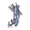
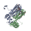
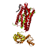
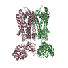
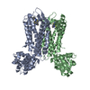
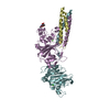
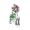
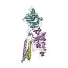
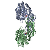
Entry Database : PDB / ID : 4rwsTitle Crystal structure of CXCR4 and viral chemokine antagonist vMIP-II complex (PSI Community Target) C-X-C chemokine receptor type 4/Endolysin chimeric protein Viral macrophage inflammatory protein 2 Keywords / / / / / / / / / / / / / Function / homology Function Domain/homology Component
/ / / / / / / / / / / / / / / / / / / / / / / / / / / / / / / / / / / / / / / / / / / / / / / / / / / / / / / / / / / / / / / / / / / / / / / / / / / / / / / / / / / / / / / / / / / / / / / / / / / / / / / / / / Biological species Homo sapiens (human)Method / / / Resolution : 3.1 Å Authors Qin, L. / Kufareva, I. / Holden, L. / Wang, C. / Zheng, Y. / Wu, H. / Fenalti, G. / Han, G.W. / Cherezov, V. / Abagyan, R. ...Qin, L. / Kufareva, I. / Holden, L. / Wang, C. / Zheng, Y. / Wu, H. / Fenalti, G. / Han, G.W. / Cherezov, V. / Abagyan, R. / Stevens, R.C. / Handel, T.M. / GPCR Network (GPCR) Journal : Science / Year : 2015Title : Structural biology. Crystal structure of the chemokine receptor CXCR4 in complex with a viral chemokine.Authors : Qin, L. / Kufareva, I. / Holden, L.G. / Wang, C. / Zheng, Y. / Zhao, C. / Fenalti, G. / Wu, H. / Han, G.W. / Cherezov, V. / Abagyan, R. / Stevens, R.C. / Handel, T.M. History Deposition Dec 5, 2014 Deposition site / Processing site Revision 1.0 Feb 11, 2015 Provider / Type Revision 1.1 Feb 25, 2015 Group Revision 1.2 Apr 15, 2015 Group Revision 1.3 Aug 2, 2017 Group / Source and taxonomy / Category / softwareRevision 1.4 Nov 22, 2017 Group / Category / Item Revision 1.5 Sep 20, 2023 Group / Database references / Refinement descriptionCategory chem_comp_atom / chem_comp_bond ... chem_comp_atom / chem_comp_bond / database_2 / pdbx_initial_refinement_model / struct_ref_seq_dif Item / _database_2.pdbx_database_accession / _struct_ref_seq_dif.detailsRevision 1.6 Nov 20, 2024 Group / Category / pdbx_modification_feature
Show all Show less
 Movie
Movie Controller

 Yorodumi
 Open data
 Basic information
 Components
 Keywords
 Function and homology information
 Homo sapiens (human)
 Enterobacteria phage T4 (virus)
 X-RAY DIFFRACTION /
  Authors
 Citation
 Structure visualization
 Downloads & links
 Other downloads
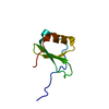


 PDBj
PDBj


 Assembly

 Spodoptera frugiperda (fall armyworm) / Strain (production host): sf9 / References: UniProt: P61073, UniProt: P00720, lysozyme
 Sample preparation
 Processing