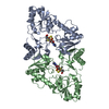
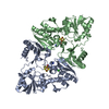
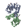
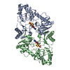
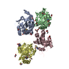
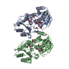
- PDB-3lzc: Crystal structure of Dph2 from Pyrococcus horikoshii -
+
Open data
ID or keywords:
Loading...
-
Basic information
Entry
Database: PDB / ID: 3lzc
Title
Crystal structure of Dph2 from Pyrococcus horikoshii
Components
Dph2
Keywords
BIOSYNTHETIC PROTEIN / Diphthamide biosynthesis / radical SAM enzyme / gene triplication
Function / homology
Function and homology information
S-adenosylmethionine catabolic process / 2-(3-amino-3-carboxypropyl)histidine synthase / 2-(3-amino-3-carboxypropyl)histidine synthase activity / protein histidyl modification to diphthamide / transferase activity, transferring alkyl or aryl (other than methyl) groups / 4 iron, 4 sulfur cluster binding / metal ion binding Similarity search - Function
Mass: 18.015 Da / Num. of mol.: 155 / Source method: isolated from a natural source / Formula: H2O
-
Experimental details
-
Experiment
Experiment
Method: X-RAY DIFFRACTION / Number of used crystals: 1
-
Sample preparation
Crystal
Density Matthews: 2.3 Å3/Da / Density % sol: 46.58 %
Crystal grow
Temperature: 291 K / Method: vapor diffusion, hanging drop / pH: 5.3 Details: 8% PEG 4000, 0.1 M ammonium acetate, 0.05 M sodium citrate, 0.2 M KCl, 2% ethylene glycol, pH 5.3, vapor diffusion, hanging drop, temperature 291K
-
Data collection
Diffraction
ID
Mean temperature (K)
Crystal-ID
1
100
1
2
100
1
Diffraction source
Source
Site
Beamline
ID
Wavelength (Å)
SYNCHROTRON
APS
24-ID-C
1
0.97922
SYNCHROTRON
APS
24-ID-E
2
0.97918
Detector
Type
ID
Detector
Date
ADSC QUANTUM 315
1
CCD
Apr 21, 2008
ADSC QUANTUM 315
2
CCD
Aug 5, 2008
Radiation
ID
Protocol
Monochromatic (M) / Laue (L)
Scattering type
Wavelength-ID
1
SINGLEWAVELENGTH
M
x-ray
1
2
SINGLEWAVELENGTH
M
x-ray
1
Radiation wavelength
ID
Wavelength (Å)
Relative weight
1
0.97922
1
2
0.97918
1
Reflection
Redundancy: 3.8 % / Av σ(I) over netI: 24.38 / Number: 196000 / Rmerge(I) obs: 0.05 / Χ2: 1.31 / D res high: 2.5 Å / D res low: 50 Å / Num. obs: 51326 / % possible obs: 99.7
In the structure databanks used in Yorodumi, some data are registered as the other names, "COVID-19 virus" and "2019-nCoV". Here are the details of the virus and the list of structure data.
Jan 31, 2019. EMDB accession codes are about to change! (news from PDBe EMDB page)
EMDB accession codes are about to change! (news from PDBe EMDB page)
The allocation of 4 digits for EMDB accession codes will soon come to an end. Whilst these codes will remain in use, new EMDB accession codes will include an additional digit and will expand incrementally as the available range of codes is exhausted. The current 4-digit format prefixed with “EMD-” (i.e. EMD-XXXX) will advance to a 5-digit format (i.e. EMD-XXXXX), and so on. It is currently estimated that the 4-digit codes will be depleted around Spring 2019, at which point the 5-digit format will come into force.
The EM Navigator/Yorodumi systems omit the EMD- prefix.
Related info.:Q: What is EMD? / ID/Accession-code notation in Yorodumi/EM Navigator
Yorodumi is a browser for structure data from EMDB, PDB, SASBDB, etc.
This page is also the successor to EM Navigator detail page, and also detail information page/front-end page for Omokage search.
The word "yorodu" (or yorozu) is an old Japanese word meaning "ten thousand". "mi" (miru) is to see.
Related info.:EMDB / PDB / SASBDB / Comparison of 3 databanks / Yorodumi Search / Aug 31, 2016. New EM Navigator & Yorodumi / Yorodumi Papers / Jmol/JSmol / Function and homology information / Changes in new EM Navigator and Yorodumi
 Movie
Movie Controller


 Open data
 Basic information
 Components
 Keywords
 Function and homology information
 Pyrococcus horikoshii (archaea)
 X-RAY DIFFRACTION /
  Authors
 Citation
 Structure visualization
 Downloads & links
 Other downloads
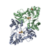


 PDBj
PDBj Assembly
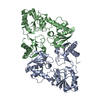

 Mass: 18.015 Da / Num. of mol.: 155 / Source method: isolated from a natural source / Formula: H2O
 Sample preparation
 Processing