 Movie
Movie Controller

[English] 日本語
 Yorodumi
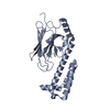
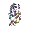
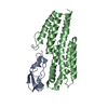
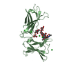
- PDB-3lx7: Crystal structure of a Novel Tudor domain-containing protein SGF29 -
+ Open data

- Basic information
| Entry | Database: PDB / ID: 3lx7 | ||||||
|---|---|---|---|---|---|---|---|
| Title | Crystal structure of a Novel Tudor domain-containing protein SGF29 | ||||||
 Components Components | SAGA-associated factor 29 homolog | ||||||
 Keywords Keywords | TRANSCRIPTION REGULATOR / SAGA / Tudor / Nucleus / Transcription / Transcription regulation | ||||||
| Function / homology |  Function and homology information Function and homology informationmethylation-dependent protein binding / SAGA-type complex / histone H3K36me3 reader activity / establishment of protein localization to chromatin / ATAC complex / histone H3K4me3 reader activity / SAGA complex / negative regulation of DNA repair / Formation of WDR5-containing histone-modifying complexes / regulation of RNA splicing ...methylation-dependent protein binding / SAGA-type complex / histone H3K36me3 reader activity / establishment of protein localization to chromatin / ATAC complex / histone H3K4me3 reader activity / SAGA complex / negative regulation of DNA repair / Formation of WDR5-containing histone-modifying complexes / regulation of RNA splicing / regulation of embryonic development / regulation of cell division / regulation of DNA repair / response to endoplasmic reticulum stress / transcription initiation-coupled chromatin remodeling / mitotic spindle / HATs acetylate histones / regulation of cell cycle / DNA repair / regulation of transcription by RNA polymerase II / regulation of DNA-templated transcription / positive regulation of DNA-templated transcription / negative regulation of transcription by RNA polymerase II / nucleoplasm Similarity search - Function | ||||||
| Biological species |  Homo sapiens (human) Homo sapiens (human) | ||||||
| Method |  X-RAY DIFFRACTION / SYNCHROTRON / MOLECULAR REPLACEMENT / Resolution: 1.78 Å X-RAY DIFFRACTION / SYNCHROTRON / MOLECULAR REPLACEMENT / Resolution: 1.78 Å | ||||||
 Authors Authors | Bian, C.B. / Xu, C. / Tempel, W. / Lam, R. / Bountra, C. / Arrowsmith, C.H. / Weigelt, J. / Edwards, A.M. / Bochkarev, A. / Min, J. | ||||||
 Citation Citation | Journal: Embo J. / Year: 2011 Title: Sgf29 binds histone H3K4me2/3 and is required for SAGA complex recruitment and histone H3 acetylation. Authors: Bian, C. / Xu, C. / Ruan, J. / Lee, K.K. / Burke, T.L. / Tempel, W. / Barsyte, D. / Li, J. / Wu, M. / Zhou, B.O. / Fleharty, B.E. / Paulson, A. / Allali-Hassani, A. / Zhou, J.Q. / Mer, G. / ...Authors: Bian, C. / Xu, C. / Ruan, J. / Lee, K.K. / Burke, T.L. / Tempel, W. / Barsyte, D. / Li, J. / Wu, M. / Zhou, B.O. / Fleharty, B.E. / Paulson, A. / Allali-Hassani, A. / Zhou, J.Q. / Mer, G. / Grant, P.A. / Workman, J.L. / Zang, J. / Min, J. | ||||||
| History |
|
- Structure visualization
| Structure viewer | Molecule: MolmilJmol/JSmol |
|---|
- Downloads & links
-Download
| PDBx/mmCIF format | 3lx7.cif.gz | 74.4 KB | Display | PDBx/mmCIF format |
|---|---|---|---|---|
| PDB format | pdb3lx7.ent.gz | 53.4 KB | Display | PDB format |
| PDBx/mmJSON format | 3lx7.json.gz | Tree view | PDBx/mmJSON format | |
| Others |  Other downloads Other downloads |
-Validation report
| Arichive directory | https://data.pdbj.org/pub/pdb/validation_reports/lx/3lx7ftp://data.pdbj.org/pub/pdb/validation_reports/lx/3lx7 | HTTPS FTP |
|---|
-Related structure data
| Related structure data | 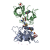 3me9SC  3meaC  3metC 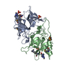 3meuC  3mevC 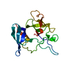 3mewC 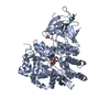 3mp1C 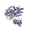 3mp6C 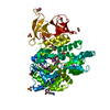 3mp8C S: Starting model for refinement C: citing same article ( |
|---|---|
| Similar structure data |


-Links
 PDBj
PDBj- Assembly
| Deposited unit | 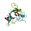
| ||||||||
|---|---|---|---|---|---|---|---|---|---|
| 1 | 
| ||||||||
| Unit cell |
|
-Components
| #1: Protein | Mass: 19750.080 Da / Num. of mol.: 1 / Fragment: Residues 138-293 Source method: isolated from a genetically manipulated source Source: (gene. exp.) Homo sapiens (human) / Gene: CCDC101, SGF29 / Plasmid: pET28-MHL / Production host:  | ||
|---|---|---|---|
| #2: Chemical | ChemComp-SO4 /   Mass: 96.063 Da / Num. of mol.: 1 / Source method: obtained synthetically / Formula: SO4 Mass: 96.063 Da / Num. of mol.: 1 / Source method: obtained synthetically / Formula: SO4 | ||
| #3: Chemical | ChemComp-UNX /   Num. of mol.: 4 / Source method: obtained synthetically Num. of mol.: 4 / Source method: obtained synthetically#4: Water | ChemComp-HOH / |  Mass: 18.015 Da / Num. of mol.: 45 / Source method: isolated from a natural source / Formula: H2O Mass: 18.015 Da / Num. of mol.: 45 / Source method: isolated from a natural source / Formula: H2O |
-Experimental details
-Experiment
| Experiment | Method: X-RAY DIFFRACTION |
|---|
- Sample preparation
| Crystal | Density Matthews: 1.93 Å3/Da / Density % sol: 36.34 % / Mosaicity: 0.432 ° |
|---|---|
| Crystal grow | Temperature: 291 K / Method: vapor diffusion, sitting drop / pH: 5.6 Details: 0.1M Sodium citrate tribasic dihydrate pH 5.6, 0.2M potassium sodium tartrate, 2M Ammonium sulfate, VAPOR DIFFUSION, SITTING DROP, temperature 291K |
-Data collection
| Diffraction | Mean temperature: 100 K | |||||||||||||||||||||||||||||||||||||||||||||||||||||||||||||||||||||||||||||||||||||||||||||||||||||||||||||||||||||||||||||||||||||||||||||||||||
|---|---|---|---|---|---|---|---|---|---|---|---|---|---|---|---|---|---|---|---|---|---|---|---|---|---|---|---|---|---|---|---|---|---|---|---|---|---|---|---|---|---|---|---|---|---|---|---|---|---|---|---|---|---|---|---|---|---|---|---|---|---|---|---|---|---|---|---|---|---|---|---|---|---|---|---|---|---|---|---|---|---|---|---|---|---|---|---|---|---|---|---|---|---|---|---|---|---|---|---|---|---|---|---|---|---|---|---|---|---|---|---|---|---|---|---|---|---|---|---|---|---|---|---|---|---|---|---|---|---|---|---|---|---|---|---|---|---|---|---|---|---|---|---|---|---|---|---|---|
| Diffraction source | Source: SYNCHROTRON / Site: CHESS  / Beamline: A1 / Wavelength: 0.978 Å / Beamline: A1 / Wavelength: 0.978 Å | |||||||||||||||||||||||||||||||||||||||||||||||||||||||||||||||||||||||||||||||||||||||||||||||||||||||||||||||||||||||||||||||||||||||||||||||||||
| Detector | Type: ADSC QUANTUM 210 / Detector: CCD / Date: May 3, 2009 | |||||||||||||||||||||||||||||||||||||||||||||||||||||||||||||||||||||||||||||||||||||||||||||||||||||||||||||||||||||||||||||||||||||||||||||||||||
| Radiation | Protocol: SINGLE WAVELENGTH / Monochromatic (M) / Laue (L): M / Scattering type: x-ray | |||||||||||||||||||||||||||||||||||||||||||||||||||||||||||||||||||||||||||||||||||||||||||||||||||||||||||||||||||||||||||||||||||||||||||||||||||
| Radiation wavelength | Wavelength: 0.978 Å / Relative weight: 1 | |||||||||||||||||||||||||||||||||||||||||||||||||||||||||||||||||||||||||||||||||||||||||||||||||||||||||||||||||||||||||||||||||||||||||||||||||||
| Reflection | Resolution: 1.77→50 Å / Num. obs: 13275 / % possible obs: 91.1 % / Redundancy: 3.3 % / Rmerge(I) obs: 0.071 / Rrim(I) all: 0.071 / Χ2: 2.367 / Net I/av σ(I): 26.236 / Net I/σ(I): 12.5 | |||||||||||||||||||||||||||||||||||||||||||||||||||||||||||||||||||||||||||||||||||||||||||||||||||||||||||||||||||||||||||||||||||||||||||||||||||
| Reflection shell |
|
- Processing
| Software |
| |||||||||||||||||||||||||||||||||||||||||||||||||||||||||||||||||||||||||||||||||||||||||||||||||||||||||||||||||||||||||||||||||||||||||||||||||||||||||||||||||||||||||||||||||||||||||||||
|---|---|---|---|---|---|---|---|---|---|---|---|---|---|---|---|---|---|---|---|---|---|---|---|---|---|---|---|---|---|---|---|---|---|---|---|---|---|---|---|---|---|---|---|---|---|---|---|---|---|---|---|---|---|---|---|---|---|---|---|---|---|---|---|---|---|---|---|---|---|---|---|---|---|---|---|---|---|---|---|---|---|---|---|---|---|---|---|---|---|---|---|---|---|---|---|---|---|---|---|---|---|---|---|---|---|---|---|---|---|---|---|---|---|---|---|---|---|---|---|---|---|---|---|---|---|---|---|---|---|---|---|---|---|---|---|---|---|---|---|---|---|---|---|---|---|---|---|---|---|---|---|---|---|---|---|---|---|---|---|---|---|---|---|---|---|---|---|---|---|---|---|---|---|---|---|---|---|---|---|---|---|---|---|---|---|---|---|---|---|---|
| Refinement | Method to determine structure: MOLECULAR REPLACEMENT Starting model: PDB ENTRY 3ME9 Resolution: 1.78→25.09 Å / Cor.coef. Fo:Fc: 0.94 / Cor.coef. Fo:Fc free: 0.911 / SU B: 6.169 / SU ML: 0.089 / Cross valid method: THROUGHOUT / ESU R: 0.149 / ESU R Free: 0.14 / Stereochemistry target values: MAXIMUM LIKELIHOOD / Details: HYDROGENS HAVE BEEN ADDED IN THE RIDING POSITIONS
| |||||||||||||||||||||||||||||||||||||||||||||||||||||||||||||||||||||||||||||||||||||||||||||||||||||||||||||||||||||||||||||||||||||||||||||||||||||||||||||||||||||||||||||||||||||||||||||
| Solvent computation | Ion probe radii: 0.8 Å / Shrinkage radii: 0.8 Å / VDW probe radii: 1.4 Å / Solvent model: MASK | |||||||||||||||||||||||||||||||||||||||||||||||||||||||||||||||||||||||||||||||||||||||||||||||||||||||||||||||||||||||||||||||||||||||||||||||||||||||||||||||||||||||||||||||||||||||||||||
| Displacement parameters | Biso mean: 23.679 Å2
| |||||||||||||||||||||||||||||||||||||||||||||||||||||||||||||||||||||||||||||||||||||||||||||||||||||||||||||||||||||||||||||||||||||||||||||||||||||||||||||||||||||||||||||||||||||||||||||
| Refinement step | Cycle: LAST / Resolution: 1.78→25.09 Å
| |||||||||||||||||||||||||||||||||||||||||||||||||||||||||||||||||||||||||||||||||||||||||||||||||||||||||||||||||||||||||||||||||||||||||||||||||||||||||||||||||||||||||||||||||||||||||||||
| Refine LS restraints |
| |||||||||||||||||||||||||||||||||||||||||||||||||||||||||||||||||||||||||||||||||||||||||||||||||||||||||||||||||||||||||||||||||||||||||||||||||||||||||||||||||||||||||||||||||||||||||||||
| LS refinement shell | Refine-ID: X-RAY DIFFRACTION / Total num. of bins used: 20
| |||||||||||||||||||||||||||||||||||||||||||||||||||||||||||||||||||||||||||||||||||||||||||||||||||||||||||||||||||||||||||||||||||||||||||||||||||||||||||||||||||||||||||||||||||||||||||||
| Refinement TLS params. | Method: refined / Origin x: 12.1841 Å / Origin y: 0.794 Å / Origin z: 18.4979 Å
|
