 Movie
Movie Controller


+ Open data

- Basic information
| Entry | Database: PDB / ID: 1qx5 | ||||||
|---|---|---|---|---|---|---|---|
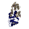
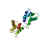
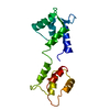
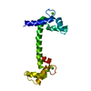
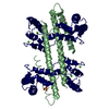
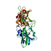
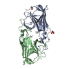
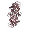
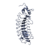
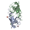
| Title | Crystal structure of apoCalmodulin | ||||||
 Components Components | Calmodulin | ||||||
 Keywords Keywords | SIGNALING PROTEIN / apoCalmodulin / domain swap / dimer / EF hands / calcium binding protein | ||||||
| Function / homology |  Function and homology information Function and homology informationregulation of store-operated calcium channel activity / : / : / regulation of response to tumor cell / positive regulation of autophagic cell death / DAPK1-calmodulin complex / : / : / : / : ...regulation of store-operated calcium channel activity / : / : / regulation of response to tumor cell / positive regulation of autophagic cell death / DAPK1-calmodulin complex / : / : / : / : / : / transporter inhibitor activity / : / type 3 metabotropic glutamate receptor binding / establishment of protein localization to membrane / positive regulation of DNA binding / negative regulation of high voltage-gated calcium channel activity / response to corticosterone / negative regulation of ryanodine-sensitive calcium-release channel activity / organelle localization by membrane tethering / : / autophagosome membrane docking / regulation of synaptic vesicle exocytosis / negative regulation of calcium ion export across plasma membrane / regulation of ryanodine-sensitive calcium-release channel activity / regulation of cardiac muscle cell action potential / presynaptic endocytosis / calcineurin-mediated signaling / nitric-oxide synthase binding / regulation of cell communication by electrical coupling involved in cardiac conduction / adenylate cyclase binding / protein phosphatase activator activity / regulation of synaptic vesicle endocytosis / detection of calcium ion / postsynaptic cytosol / regulation of cardiac muscle contraction / cell surface receptor signaling pathway via JAK-STAT / catalytic complex / phosphatidylinositol 3-kinase binding / positive regulation of nitric-oxide synthase activity / activation of adenylate cyclase activity / calcium channel inhibitor activity / presynaptic cytosol / cellular response to interferon-beta / enzyme regulator activity / regulation of release of sequestered calcium ion into cytosol by sarcoplasmic reticulum / titin binding / regulation of cardiac muscle contraction by regulation of the release of sequestered calcium ion / regulation of calcium-mediated signaling / voltage-gated potassium channel complex / calcium channel complex / potassium ion transmembrane transport / regulation of heart rate / calyx of Held / nitric-oxide synthase regulator activity / adenylate cyclase activator activity / regulation of cytokinesis / protein serine/threonine kinase activator activity / spindle microtubule / positive regulation of receptor signaling pathway via JAK-STAT / sarcomere / response to amphetamine / calcium channel regulator activity / calcium-mediated signaling / response to calcium ion / cellular response to type II interferon / G2/M transition of mitotic cell cycle / Schaffer collateral - CA1 synapse / spindle pole / disordered domain specific binding / calcium-dependent protein binding / protein autophosphorylation / myelin sheath / synaptic vesicle membrane / growth cone / sperm midpiece / vesicle / transmembrane transporter binding / neuron projection / positive regulation of apoptotic process / protein domain specific binding / calcium ion binding / centrosome / protein kinase binding / chromatin / protein-containing complex / mitochondrion / nucleoplasm / membrane / nucleus / plasma membrane / cytoplasm / cytosol Similarity search - Function | ||||||
| Biological species |  | ||||||
| Method |  X-RAY DIFFRACTION / SYNCHROTRON / MIR / Resolution: 2.54 Å X-RAY DIFFRACTION / SYNCHROTRON / MIR / Resolution: 2.54 Å | ||||||
 Authors Authors | Schumacher, M.A. / Crum, M. / Miller, M.C. | ||||||
 Citation Citation | Journal: STRUCTURE / Year: 2004 Title: Crystal structures of apocalmodulin and an apocalmodulin/SK potassium channel gating domain complex. Authors: Schumacher, M.A. / Crum, M. / Miller, M.C. | ||||||
| History |
|
- Structure visualization
| Structure viewer | Molecule: MolmilJmol/JSmol |
|---|
- Downloads & links
-Download
| PDBx/mmCIF format | 1qx5.cif.gz | 232.4 KB | Display | PDBx/mmCIF format |
|---|---|---|---|---|
| PDB format | pdb1qx5.ent.gz | 191.9 KB | Display | PDB format |
| PDBx/mmJSON format | 1qx5.json.gz | Tree view | PDBx/mmJSON format | |
| Others |  Other downloads Other downloads |
-Validation report
| Arichive directory | https://data.pdbj.org/pub/pdb/validation_reports/qx/1qx5ftp://data.pdbj.org/pub/pdb/validation_reports/qx/1qx5 | HTTPS FTP |
|---|
-Related structure data


-Links
 PDBj
PDBj

- Assembly
| Deposited unit | 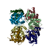
| ||||||||
|---|---|---|---|---|---|---|---|---|---|
| 1 | 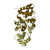
| ||||||||
| 2 | 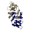
| ||||||||
| 3 | 
| ||||||||
| 4 | 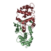
| ||||||||
| Unit cell |
| ||||||||
| Details | apoCaM forms a domain swapped dimer: there are four, essentially identical dimers in the ASU |
-Components
| #1: Protein | Mass: 16721.350 Da / Num. of mol.: 8 Source method: isolated from a genetically manipulated source Source: (gene. exp.) Gene: CALM1, CAM1, CALM, CAM, CALM2, CAM2, CAMB, CALM3, CAM3, CAMC Plasmid: pET23b / Species (production host): Escherichia coli / Production host:  #2: Water | ChemComp-HOH / |  Mass: 18.015 Da / Num. of mol.: 175 / Source method: isolated from a natural source / Formula: H2O Mass: 18.015 Da / Num. of mol.: 175 / Source method: isolated from a natural source / Formula: H2O |
|---|
-Experimental details
-Experiment
| Experiment | Method: X-RAY DIFFRACTION / Number of used crystals: 1 |
|---|
- Sample preparation
| Crystal | Density Matthews: 3.59 Å3/Da / Density % sol: 65.71 % |
|---|---|
| Crystal grow | Temperature: 298 K / Method: vapor diffusion, hanging drop / pH: 6.5 Details: Citrate, 150 mM NaCl, Hepes , pH 6.5, VAPOR DIFFUSION, HANGING DROP, temperature 298K |
-Data collection
| Diffraction | Mean temperature: 100 K |
|---|---|
| Diffraction source | Source: SYNCHROTRON / Site: SSRL  / Beamline: BL9-1 / Wavelength: 0.989 Å / Beamline: BL9-1 / Wavelength: 0.989 Å |
| Detector | Type: ADSC QUANTUM 4 / Detector: CCD / Date: Feb 12, 2003 |
| Radiation | Monochromator: graphite / Protocol: SINGLE WAVELENGTH / Monochromatic (M) / Laue (L): M / Scattering type: x-ray |
| Radiation wavelength | Wavelength: 0.989 Å / Relative weight: 1 |
| Reflection | Resolution: 2.54→66.4 Å / Num. all: 61810 / Num. obs: 61810 / % possible obs: 98.5 % / Observed criterion σ(F): 0 / Observed criterion σ(I): 0 / Biso Wilson estimate: 75.4 Å2 / Rsym value: 0.047 / Net I/σ(I): 10.2 |
| Reflection shell | Resolution: 2.54→2.7 Å / % possible all: 98.6 |
- Processing
| Software |
| |||||||||||||||||||||||||
|---|---|---|---|---|---|---|---|---|---|---|---|---|---|---|---|---|---|---|---|---|---|---|---|---|---|---|
| Refinement | Method to determine structure: MIR / Resolution: 2.54→66.39 Å / Rfactor Rfree error: 0.005 / Isotropic thermal model: RESTRAINED / Cross valid method: THROUGHOUT / σ(F): 0 / Stereochemistry target values: Engh & Huber
| |||||||||||||||||||||||||
| Solvent computation | Solvent model: FLAT MODEL / Bsol: 60.8149 Å2 / ksol: 0.374381 e/Å3 | |||||||||||||||||||||||||
| Displacement parameters | Biso mean: 71.1 Å2
| |||||||||||||||||||||||||
| Refine analyze | Luzzati coordinate error free: 0.48 Å / Luzzati sigma a free: 0.51 Å | |||||||||||||||||||||||||
| Refinement step | Cycle: LAST / Resolution: 2.54→66.39 Å
| |||||||||||||||||||||||||
| Refine LS restraints |
| |||||||||||||||||||||||||
| LS refinement shell | Resolution: 2.54→2.7 Å / Rfactor Rfree error: 0.022 / Total num. of bins used: 6
| |||||||||||||||||||||||||
| Xplor file |
|
