 Movie
Movie Controller

[English] 日本語
 Yorodumi
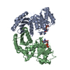
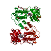
- PDB-1g4y: 1.60 A CRYSTAL STRUCTURE OF THE GATING DOMAIN FROM SMALL CONDUCTA... -
+ Open data

- Basic information
| Entry | Database: PDB / ID: 1g4y | ||||||
|---|---|---|---|---|---|---|---|
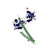
| Title | 1.60 A CRYSTAL STRUCTURE OF THE GATING DOMAIN FROM SMALL CONDUCTANCE POTASSIUM CHANNEL COMPLEXED WITH CALCIUM-CALMODULIN | ||||||
 Components Components |
| ||||||
 Keywords Keywords | SIGNALING PROTEIN / small-conductance calcium-activated potassium channel / calmodulin / calmodulin binding domain (CaMBD) / channel gating | ||||||
| Function / homology |  Function and homology information Function and homology informationCa2+ activated K+ channels / regulation of store-operated calcium channel activity / : / : / small conductance calcium-activated potassium channel activity / membrane repolarization during atrial cardiac muscle cell action potential / regulation of response to tumor cell / positive regulation of autophagic cell death / DAPK1-calmodulin complex / : ...Ca2+ activated K+ channels / regulation of store-operated calcium channel activity / : / : / small conductance calcium-activated potassium channel activity / membrane repolarization during atrial cardiac muscle cell action potential / regulation of response to tumor cell / positive regulation of autophagic cell death / DAPK1-calmodulin complex / : / : / : / : / calcium-activated potassium channel activity / : / transporter inhibitor activity / positive regulation of potassium ion transport / : / type 3 metabotropic glutamate receptor binding / inward rectifier potassium channel activity / establishment of protein localization to membrane / regulation of potassium ion transmembrane transport / positive regulation of DNA binding / negative regulation of high voltage-gated calcium channel activity / response to corticosterone / negative regulation of ryanodine-sensitive calcium-release channel activity / organelle localization by membrane tethering / : / autophagosome membrane docking / regulation of synaptic vesicle exocytosis / negative regulation of calcium ion export across plasma membrane / regulation of ryanodine-sensitive calcium-release channel activity / regulation of cardiac muscle cell action potential / presynaptic endocytosis / smooth endoplasmic reticulum / calcineurin-mediated signaling / nitric-oxide synthase binding / regulation of cell communication by electrical coupling involved in cardiac conduction / alpha-actinin binding / adenylate cyclase binding / protein phosphatase activator activity / regulation of synaptic vesicle endocytosis / monoatomic ion channel activity / regulation of neuronal synaptic plasticity / detection of calcium ion / postsynaptic cytosol / regulation of cardiac muscle contraction / cell surface receptor signaling pathway via JAK-STAT / catalytic complex / positive regulation of nitric-oxide synthase activity / phosphatidylinositol 3-kinase binding / activation of adenylate cyclase activity / calcium channel inhibitor activity / presynaptic cytosol / cellular response to interferon-beta / enzyme regulator activity / regulation of release of sequestered calcium ion into cytosol by sarcoplasmic reticulum / titin binding / monoatomic ion transport / regulation of cardiac muscle contraction by regulation of the release of sequestered calcium ion / regulation of calcium-mediated signaling / voltage-gated potassium channel complex / calcium channel complex / T-tubule / potassium ion transmembrane transport / regulation of heart rate / calyx of Held / nitric-oxide synthase regulator activity / adenylate cyclase activator activity / protein serine/threonine kinase activator activity / regulation of cytokinesis / spindle microtubule / positive regulation of receptor signaling pathway via JAK-STAT / response to amphetamine / sarcomere / calcium channel regulator activity / potassium ion transport / calcium-mediated signaling / sarcolemma / response to calcium ion / cellular response to type II interferon / modulation of chemical synaptic transmission / G2/M transition of mitotic cell cycle / Schaffer collateral - CA1 synapse / Z disc / spindle pole / disordered domain specific binding / calcium-dependent protein binding / protein autophosphorylation / myelin sheath / synaptic vesicle membrane / growth cone / sperm midpiece / vesicle / dendritic spine / transmembrane transporter binding / calmodulin binding / postsynaptic membrane / neuron projection / positive regulation of apoptotic process Similarity search - Function | ||||||
| Biological species |  | ||||||
| Method |  X-RAY DIFFRACTION / SYNCHROTRON / MIR / Resolution: 1.6 Å X-RAY DIFFRACTION / SYNCHROTRON / MIR / Resolution: 1.6 Å | ||||||
 Authors Authors | Schumacher, M.A. / Rivard, A. / Bachinger, H.P. / Adelman, J.P. | ||||||
 Citation Citation | Journal: Nature / Year: 2001 Title: Structure of the gating domain of a Ca2+-activated K+ channel complexed with Ca2+/calmodulin. Authors: Schumacher, M.A. / Rivard, A.F. / Bachinger, H.P. / Adelman, J.P. | ||||||
| History |
|
- Structure visualization
| Structure viewer | Molecule: MolmilJmol/JSmol |
|---|
- Downloads & links
-Download
| PDBx/mmCIF format | 1g4y.cif.gz | 64.5 KB | Display | PDBx/mmCIF format |
|---|---|---|---|---|
| PDB format | pdb1g4y.ent.gz | 47 KB | Display | PDB format |
| PDBx/mmJSON format | 1g4y.json.gz | Tree view | PDBx/mmJSON format | |
| Others |  Other downloads Other downloads |
-Validation report
| Arichive directory | https://data.pdbj.org/pub/pdb/validation_reports/g4/1g4yftp://data.pdbj.org/pub/pdb/validation_reports/g4/1g4y | HTTPS FTP |
|---|
-Related structure data
| Similar structure data |
|---|


-Links
 PDBj
PDBj


- Assembly
| Deposited unit | 
| |||||||||
|---|---|---|---|---|---|---|---|---|---|---|
| 1 | 
| |||||||||
| Unit cell |
| |||||||||
| Components on special symmetry positions |
| |||||||||
| Details | the relevant dimer is generated by crystallographic symmetry. |
-Components
| #1: Protein | Mass: 12105.248 Da / Num. of mol.: 1 / Fragment: CALMODULIN-BINDING DOMAIN Source method: isolated from a genetically manipulated source Source: (gene. exp.)  | ||
|---|---|---|---|
| #2: Protein | Mass: 16721.350 Da / Num. of mol.: 1 Source method: isolated from a genetically manipulated source Source: (gene. exp.) | ||
| #3: Chemical | ChemComp-SO4 /   Mass: 96.063 Da / Num. of mol.: 1 / Source method: obtained synthetically / Formula: SO4 Mass: 96.063 Da / Num. of mol.: 1 / Source method: obtained synthetically / Formula: SO4 | ||
| #4: Chemical |   Mass: 40.078 Da / Num. of mol.: 2 / Source method: obtained synthetically / Formula: Ca Mass: 40.078 Da / Num. of mol.: 2 / Source method: obtained synthetically / Formula: Ca#5: Water | ChemComp-HOH / |  Mass: 18.015 Da / Num. of mol.: 199 / Source method: isolated from a natural source / Formula: H2O Mass: 18.015 Da / Num. of mol.: 199 / Source method: isolated from a natural source / Formula: H2O |
-Experimental details
-Experiment
| Experiment | Method: X-RAY DIFFRACTION / Number of used crystals: 1 |
|---|
- Sample preparation
| Crystal | Density Matthews: 2.9 Å3/Da / Density % sol: 57.57 % | ||||||||||||||||||||||||||||||||||||||||||
|---|---|---|---|---|---|---|---|---|---|---|---|---|---|---|---|---|---|---|---|---|---|---|---|---|---|---|---|---|---|---|---|---|---|---|---|---|---|---|---|---|---|---|---|
| Crystal grow | Temperature: 298 K / Method: vapor diffusion, hanging drop / pH: 5.6 Details: ammonium sulphate, lithium sulphate, citrate, pH 5.6, VAPOR DIFFUSION, HANGING DROP, temperature 298K | ||||||||||||||||||||||||||||||||||||||||||
| Crystal grow | *PLUS pH: 7.5 | ||||||||||||||||||||||||||||||||||||||||||
| Components of the solutions | *PLUS
|
-Data collection
| Diffraction | Mean temperature: 110 K |
|---|---|
| Diffraction source | Source: SYNCHROTRON / Site: SSRL  / Beamline: BL9-2 / Wavelength: 1.08 Å / Beamline: BL9-2 / Wavelength: 1.08 Å |
| Detector | Type: ADSC QUANTUM 4 / Detector: CCD / Date: Dec 2, 2000 / Details: mirrors |
| Radiation | Monochromator: double-crystal Si 111 crystals / Protocol: SINGLE WAVELENGTH / Monochromatic (M) / Laue (L): M / Scattering type: x-ray |
| Radiation wavelength | Wavelength: 1.08 Å / Relative weight: 1 |
| Reflection | Resolution: 1.6→40 Å / Num. all: 115320 / Num. obs: 40694 / % possible obs: 94.1 % / Observed criterion σ(F): 0 / Observed criterion σ(I): 0 / Redundancy: 2.8 % / Biso Wilson estimate: 36 Å2 / Rmerge(I) obs: 0.06 / Rsym value: 5 / Net I/σ(I): 8.2 |
| Reflection shell | Resolution: 1.6→1.64 Å / Redundancy: 2.3 % / Rmerge(I) obs: 0.32 / Mean I/σ(I) obs: 2.5 / Num. unique all: 5200 / Rsym value: 28.9 / % possible all: 87.9 |
| Reflection | *PLUS Num. measured all: 115320 |
- Processing
| Software |
| |||||||||||||||||||||||||
|---|---|---|---|---|---|---|---|---|---|---|---|---|---|---|---|---|---|---|---|---|---|---|---|---|---|---|
| Refinement | Method to determine structure: MIR Starting model: room temp model Resolution: 1.6→40 Å / Isotropic thermal model: isotropic / Cross valid method: THROUGHOUT / σ(F): 0 / σ(I): 0 / Stereochemistry target values: engh and huber
| |||||||||||||||||||||||||
| Displacement parameters | Biso mean: 4 Å2
| |||||||||||||||||||||||||
| Refine analyze |
| |||||||||||||||||||||||||
| Refinement step | Cycle: LAST / Resolution: 1.6→40 Å
| |||||||||||||||||||||||||
| Refine LS restraints |
| |||||||||||||||||||||||||
| Software | *PLUS Name: CNS / Classification: refinement | |||||||||||||||||||||||||
| Refinement | *PLUS Lowest resolution: 40 Å / σ(F): 0 / % reflection Rfree: 10 % / Rfactor all: 0.221 | |||||||||||||||||||||||||
| Solvent computation | *PLUS | |||||||||||||||||||||||||
| Displacement parameters | *PLUS Biso mean: 4 Å2 |
