Mass: 18.015 Da / Num. of mol.: 174 / Source method: isolated from a natural source / Formula: H2O
Has protein modification
Y
Sequence details
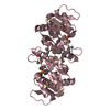
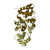
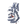
THE CONSTRUCT (RESIDUES 23-317) WAS EXPRESSED WITH A PURIFICATION TAG MGSDKIHHHHHHENLYFQG. THE TAG ...THE CONSTRUCT (RESIDUES 23-317) WAS EXPRESSED WITH A PURIFICATION TAG MGSDKIHHHHHHENLYFQG. THE TAG WAS REMOVED WITH TEV PROTEASE LEAVING ONLY A GLYCINE (0) FOLLOWED BY THE TARGET SEQUENCE.
-
Experimental details
-
Experiment
Experiment
Method: X-RAY DIFFRACTION / Number of used crystals: 1
-
Sample preparation
Crystal
Density Matthews: 3.25 Å3/Da / Density % sol: 62.16 %
Crystal grow
Temperature: 277 K / Method: vapor diffusion, sitting drop Details: 1.4M sodium citrate, 0.1 M HEPES pH 7.5, NANODROP, VAPOR DIFFUSION, SITTING DROP, temperature 277K
Resolution: 2.2→29.643 Å / Num. obs: 23862 / % possible obs: 99.9 % / Redundancy: 4 % / Biso Wilson estimate: 35.684 Å2 / Rmerge(I) obs: 0.097 / Rsym value: 0.097 / Net I/σ(I): 6.793
Reflection shell
Diffraction-ID: 1
Resolution (Å)
Redundancy (%)
Rmerge(I) obs
Mean I/σ(I) obs
Num. measured all
Num. unique all
Rsym value
% possible all
2.2-2.26
4
0.65
1.2
7014
1745
0.65
100
2.26-2.32
4
0.576
1.4
6758
1682
0.576
100
2.32-2.39
4
0.486
1.6
6584
1644
0.486
100
2.39-2.46
4
0.434
1.8
6419
1596
0.434
100
2.46-2.54
4
0.369
2.1
6241
1554
0.369
100
2.54-2.63
4
0.304
2.5
6020
1505
0.304
100
2.63-2.73
4
0.255
3.1
5815
1446
0.255
100
2.73-2.84
4
0.211
3.7
5594
1403
0.211
100
2.84-2.97
4
0.156
5
5435
1355
0.156
100
2.97-3.11
4
0.122
6.2
5092
1278
0.122
100
3.11-3.28
4
0.096
7.4
4924
1241
0.096
100
3.28-3.48
4
0.079
8.8
4644
1170
0.079
100
3.48-3.72
4
0.066
10.3
4368
1104
0.066
100
3.72-4.02
3.9
0.057
11.3
4051
1029
0.057
100
4.02-4.4
3.9
0.05
13.2
3753
962
0.05
100
4.4-4.92
3.9
0.043
14.8
3398
869
0.043
100
4.92-5.68
3.8
0.049
12.7
2981
781
0.049
99.9
5.68-6.96
3.8
0.059
11.4
2482
660
0.059
99.8
6.96-9.84
3.6
0.051
11.7
1916
536
0.051
99.6
9.84-29.65
3.3
0.042
14.3
990
302
0.042
94.3
-
Phasing
Phasing
Method: MAD
-
Processing
Software
Name
Version
Classification
NB
REFMAC
5.5.0053
refinement
PHENIX
refinement
SHELX
phasing
MolProbity
3beta29
modelbuilding
SCALA
3.2.5
datascaling
PDB_EXTRACT
3.006
dataextraction
MOSFLM
datareduction
SHELXD
phasing
autoSHARP
phasing
Refinement
Method to determine structure: MAD / Resolution: 2.2→29.643 Å / Cor.coef. Fo:Fc: 0.952 / Cor.coef. Fo:Fc free: 0.925 / Occupancy max: 1 / Occupancy min: 0.07 / SU B: 9.773 / SU ML: 0.11 / TLS residual ADP flag: LIKELY RESIDUAL / Cross valid method: THROUGHOUT / σ(F): 0 / ESU R: 0.188 / ESU R Free: 0.171 Stereochemistry target values: MAXIMUM LIKELIHOOD WITH PHASES Details: 1. HYDROGENS HAVE BEEN ADDED IN THE RIDING POSITIONS. 2. A MET-INHIBITION PROTOCOL WAS USED FOR SELENOMETHIONINE INCORPORATION DURING PROTEIN EXPRESSION. THE OCCUPANCY OF THE SE ATOMS IN THE ...Details: 1. HYDROGENS HAVE BEEN ADDED IN THE RIDING POSITIONS. 2. A MET-INHIBITION PROTOCOL WAS USED FOR SELENOMETHIONINE INCORPORATION DURING PROTEIN EXPRESSION. THE OCCUPANCY OF THE SE ATOMS IN THE MSE RESIDUES WAS REDUCED TO 0.75 FOR THE REDUCED SCATTERING POWER DUE TO PARTIAL S-MET INCORPORATION. 3. ATOM RECORDS CONTAIN RESIDUAL B FACTORS ONLY. 4. EDO MODELED IS PRESENT IN CRYO SOLUTION. 5. DENSITY FOR RESIDUES 220-229 ARE POOR.
Rfactor
Num. reflection
% reflection
Selection details
Rfree
0.229
1220
5.1 %
RANDOM
Rwork
0.189
-
-
-
obs
0.191
23827
99.84 %
-
Solvent computation
Ion probe radii: 0.8 Å / Shrinkage radii: 0.8 Å / VDW probe radii: 1.2 Å / Solvent model: MASK
In the structure databanks used in Yorodumi, some data are registered as the other names, "COVID-19 virus" and "2019-nCoV". Here are the details of the virus and the list of structure data.
Jan 31, 2019. EMDB accession codes are about to change! (news from PDBe EMDB page)
EMDB accession codes are about to change! (news from PDBe EMDB page)
The allocation of 4 digits for EMDB accession codes will soon come to an end. Whilst these codes will remain in use, new EMDB accession codes will include an additional digit and will expand incrementally as the available range of codes is exhausted. The current 4-digit format prefixed with “EMD-” (i.e. EMD-XXXX) will advance to a 5-digit format (i.e. EMD-XXXXX), and so on. It is currently estimated that the 4-digit codes will be depleted around Spring 2019, at which point the 5-digit format will come into force.
The EM Navigator/Yorodumi systems omit the EMD- prefix.
Related info.:Q: What is EMD? / ID/Accession-code notation in Yorodumi/EM Navigator
Yorodumi is a browser for structure data from EMDB, PDB, SASBDB, etc.
This page is also the successor to EM Navigator detail page, and also detail information page/front-end page for Omokage search.
The word "yorodu" (or yorozu) is an old Japanese word meaning "ten thousand". "mi" (miru) is to see.
Related info.:EMDB / PDB / SASBDB / Comparison of 3 databanks / Yorodumi Search / Aug 31, 2016. New EM Navigator & Yorodumi / Yorodumi Papers / Jmol/JSmol / Function and homology information / Changes in new EM Navigator and Yorodumi
 Movie
Movie Controller

 Yorodumi
 Open data
 Basic information
 Components
 Keywords
 Function and homology information
 Bacteroides thetaiotaomicron VPI-5482 (bacteria)
 X-RAY DIFFRACTION /
  Authors
 Citation
 Structure visualization
 Downloads & links
 Other downloads
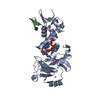

 PDBj
PDBj


 Assembly

 Mass: 62.068 Da / Num. of mol.: 1 / Source method: obtained synthetically / Formula: C2H6O2
 Mass: 18.015 Da / Num. of mol.: 174 / Source method: isolated from a natural source / Formula: H2O
 Sample preparation
 / Beamline: BL9-2 / Wavelength: 0.91162,0.97927,0.97915
 Processing