 Movie
Movie Controller

[English] 日本語
 Yorodumi
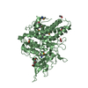
- PDB-3lew: Crystal structure of SusD-like carbohydrate binding protein (YP_0... -
+ Open data

- Basic information
| Entry | Database: PDB / ID: 3lew | ||||||
|---|---|---|---|---|---|---|---|
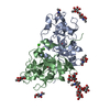
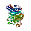
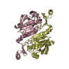
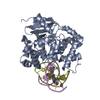
| Title | Crystal structure of SusD-like carbohydrate binding protein (YP_001298396.1) from Bacteroides vulgatus ATCC 8482 at 1.70 A resolution | ||||||
 Components Components | SusD-like carbohydrate binding protein | ||||||
 Keywords Keywords | SUGAR BINDING PROTEIN / SusD-like carbohydrate binding protein / Structural Genomics / Joint Center for Structural Genomics / JCSG / Protein Structure Initiative / PSI-2 | ||||||
| Function / homology |  Function and homology information Function and homology information | ||||||
| Biological species |  Bacteroides vulgatus (bacteria) Bacteroides vulgatus (bacteria) | ||||||
| Method |  X-RAY DIFFRACTION / SYNCHROTRON / MAD / Resolution: 1.7 Å X-RAY DIFFRACTION / SYNCHROTRON / MAD / Resolution: 1.7 Å | ||||||
 Authors Authors | Joint Center for Structural Genomics (JCSG) | ||||||
 Citation Citation | Journal: To be published Title: Crystal structure of SusD-like carbohydrate binding protein (YP_001298396.1) from Bacteroides vulgatus ATCC 8482 at 1.70 A resolution Authors: Joint Center for Structural Genomics (JCSG) | ||||||
| History |
|
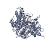
- Structure visualization
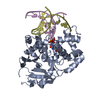
| Structure viewer | Molecule: MolmilJmol/JSmol |
|---|
- Downloads & links
-Download
| PDBx/mmCIF format | 3lew.cif.gz | 131 KB | Display | PDBx/mmCIF format |
|---|---|---|---|---|
| PDB format | pdb3lew.ent.gz | 97.6 KB | Display | PDB format |
| PDBx/mmJSON format | 3lew.json.gz | Tree view | PDBx/mmJSON format | |
| Others |  Other downloads Other downloads |
-Validation report
| Arichive directory | https://data.pdbj.org/pub/pdb/validation_reports/le/3lewftp://data.pdbj.org/pub/pdb/validation_reports/le/3lew | HTTPS FTP |
|---|
-Related structure data
| Similar structure data | |
|---|---|
| Other databases |

-Links
 PDBj
PDBj


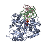
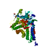
- Assembly
| Deposited unit | 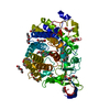
| ||||||||
|---|---|---|---|---|---|---|---|---|---|
| 1 |
| ||||||||
| Unit cell |
| ||||||||
| Components on special symmetry positions |
| ||||||||
| Details | CRYSTAL PACKING ANALYSIS SUGGESTS THE ASSIGNMENT OF A MONOMER AS THE SIGNIFICANT OLIGOMERIZATION STATE. |
-Components
| #1: Protein | Mass: 56691.859 Da / Num. of mol.: 1 / Fragment: sequence database residues 23-516 Source method: isolated from a genetically manipulated source Source: (gene. exp.) Bacteroides vulgatus (bacteria) / Strain: ATCC 8482 / DSM 1447 / NCTC 11154 / Gene: BVU_1083 / Plasmid: SpeedET / Production host: | ||||||||
|---|---|---|---|---|---|---|---|---|---|
| #2: Chemical | ChemComp-1PE /   Mass: 238.278 Da / Num. of mol.: 12 / Source method: obtained synthetically / Formula: C10H22O6 / Comment: precipitant*YM Mass: 238.278 Da / Num. of mol.: 12 / Source method: obtained synthetically / Formula: C10H22O6 / Comment: precipitant*YM#3: Chemical | ChemComp-2PE / |   Mass: 414.488 Da / Num. of mol.: 1 / Source method: obtained synthetically / Formula: C18H38O10 / Comment: precipitant*YM Mass: 414.488 Da / Num. of mol.: 1 / Source method: obtained synthetically / Formula: C18H38O10 / Comment: precipitant*YM#4: Water | ChemComp-HOH / |  Mass: 18.015 Da / Num. of mol.: 527 / Source method: isolated from a natural source / Formula: H2O Mass: 18.015 Da / Num. of mol.: 527 / Source method: isolated from a natural source / Formula: H2OHas protein modification | Y | Sequence details | THIS CONSTRUCT (RESIDUES 23-516) WAS EXPRESSED WITH A PURIFICATION TAG MGSDKIHHHHHHENLYFQG. THE TAG ...THIS CONSTRUCT (RESIDUES 23-516) WAS EXPRESSED WITH A PURIFICATI | |
-Experimental details
-Experiment
| Experiment | Method: X-RAY DIFFRACTION / Number of used crystals: 1 |
|---|
- Sample preparation
| Crystal | Density Matthews: 2.78 Å3/Da / Density % sol: 55.82 % |
|---|---|
| Crystal grow | Temperature: 277 K / Method: vapor diffusion, sitting drop / pH: 6.5 Details: 5.0000% polyethylene glycol 3000, 32.0000% polyethylene glycol 400, 0.1M MES pH 6.5, NANODROP', VAPOR DIFFUSION, SITTING DROP, temperature 277K |
-Data collection
| Diffraction | Mean temperature: 100 K | |||||||||||||||||||||||||||||||||||||||||||||||||||||||||||||||||||||||||||||
|---|---|---|---|---|---|---|---|---|---|---|---|---|---|---|---|---|---|---|---|---|---|---|---|---|---|---|---|---|---|---|---|---|---|---|---|---|---|---|---|---|---|---|---|---|---|---|---|---|---|---|---|---|---|---|---|---|---|---|---|---|---|---|---|---|---|---|---|---|---|---|---|---|---|---|---|---|---|---|
| Diffraction source | Source: SYNCHROTRON / Site: SSRL  / Beamline: BL9-2 / Wavelength: 0.91837,0.97954 / Beamline: BL9-2 / Wavelength: 0.91837,0.97954 | |||||||||||||||||||||||||||||||||||||||||||||||||||||||||||||||||||||||||||||
| Detector | Type: MARMOSAIC 325 mm CCD / Detector: CCD / Date: Nov 6, 2009 / Details: Flat collimating mirror, toroid focusing mirror | |||||||||||||||||||||||||||||||||||||||||||||||||||||||||||||||||||||||||||||
| Radiation | Monochromator: Double crystal monochromator / Protocol: MAD / Monochromatic (M) / Laue (L): M / Scattering type: x-ray | |||||||||||||||||||||||||||||||||||||||||||||||||||||||||||||||||||||||||||||
| Radiation wavelength |
| |||||||||||||||||||||||||||||||||||||||||||||||||||||||||||||||||||||||||||||
| Reflection | Resolution: 1.7→39.368 Å / Num. obs: 68815 / % possible obs: 99.5 % / Observed criterion σ(I): -3 / Biso Wilson estimate: 27.053 Å2 / Rmerge(I) obs: 0.07 / Net I/σ(I): 11.49 | |||||||||||||||||||||||||||||||||||||||||||||||||||||||||||||||||||||||||||||
| Reflection shell | Diffraction-ID: 1
|
-Phasing
| Phasing | Method: MAD |
|---|
- Processing
| Software |
| |||||||||||||||||||||||||||||||||||||||||||||||||||||||||||||||||||||||||||||||||||||
|---|---|---|---|---|---|---|---|---|---|---|---|---|---|---|---|---|---|---|---|---|---|---|---|---|---|---|---|---|---|---|---|---|---|---|---|---|---|---|---|---|---|---|---|---|---|---|---|---|---|---|---|---|---|---|---|---|---|---|---|---|---|---|---|---|---|---|---|---|---|---|---|---|---|---|---|---|---|---|---|---|---|---|---|---|---|---|
| Refinement | Method to determine structure: MAD / Resolution: 1.7→39.368 Å / Cor.coef. Fo:Fc: 0.973 / Cor.coef. Fo:Fc free: 0.966 / Occupancy max: 1 / Occupancy min: 0.22 / SU B: 3.611 / SU ML: 0.052 / TLS residual ADP flag: LIKELY RESIDUAL / Cross valid method: THROUGHOUT / σ(F): 0 / ESU R: 0.078 / ESU R Free: 0.078 Stereochemistry target values: MAXIMUM LIKELIHOOD WITH PHASES Details: 1. HYDROGENS HAVE BEEN ADDED IN THE RIDING POSITIONS. 2. ATOM RECORDS CONTAIN RESIDUAL B FACTORS ONLY. 3. A MET-INHIBITION PROTOCOL WAS USED FOR SELENOMETHIONINE INCORPORATION DURING PROTEIN ...Details: 1. HYDROGENS HAVE BEEN ADDED IN THE RIDING POSITIONS. 2. ATOM RECORDS CONTAIN RESIDUAL B FACTORS ONLY. 3. A MET-INHIBITION PROTOCOL WAS USED FOR SELENOMETHIONINE INCORPORATION DURING PROTEIN EXPRESSION. THE OCCUPANCY OF THE SE ATOMS IN THE MSE RESIDUES WAS REDUCED TO 0.75 FOR THE REDUCED SCATTERING POWER DUE TO PARTIAL S-MET INCORPORATION. 4. PEG-400 FRAGMENTS (1PE AND 2PE) FROM THE CRYOPROTECTION SOLUTION ARE MODELED.
| |||||||||||||||||||||||||||||||||||||||||||||||||||||||||||||||||||||||||||||||||||||
| Solvent computation | Ion probe radii: 0.8 Å / Shrinkage radii: 0.8 Å / VDW probe radii: 1.4 Å / Solvent model: BABINET MODEL WITH MASK | |||||||||||||||||||||||||||||||||||||||||||||||||||||||||||||||||||||||||||||||||||||
| Displacement parameters | Biso max: 70.37 Å2 / Biso mean: 20.068 Å2 / Biso min: 5.67 Å2
| |||||||||||||||||||||||||||||||||||||||||||||||||||||||||||||||||||||||||||||||||||||
| Refinement step | Cycle: LAST / Resolution: 1.7→39.368 Å
| |||||||||||||||||||||||||||||||||||||||||||||||||||||||||||||||||||||||||||||||||||||
| Refine LS restraints |
| |||||||||||||||||||||||||||||||||||||||||||||||||||||||||||||||||||||||||||||||||||||
| LS refinement shell | Resolution: 1.7→1.744 Å / Total num. of bins used: 20
| |||||||||||||||||||||||||||||||||||||||||||||||||||||||||||||||||||||||||||||||||||||
| Refinement TLS params. | Method: refined / Origin x: 58.508 Å / Origin y: 2.349 Å / Origin z: 22.912 Å
| |||||||||||||||||||||||||||||||||||||||||||||||||||||||||||||||||||||||||||||||||||||
| Refinement TLS group |
|
