 Movie
Movie Controller

[English] 日本語
 Yorodumi
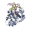
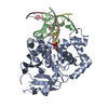
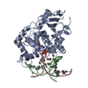
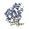
- PDB-1lww: Borohydride-trapped hOgg1 Intermediate Structure Co-Crystallized ... -
+ Open data

- Basic information
| Entry | Database: PDB / ID: 1lww | ||||||
|---|---|---|---|---|---|---|---|
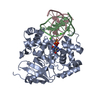
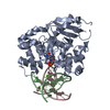
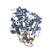
| Title | Borohydride-trapped hOgg1 Intermediate Structure Co-Crystallized with 8-bromoguanine | ||||||
 Components Components |
| ||||||
 Keywords Keywords | HYDROLASE/DNA / DNA REPAIR / DNA GLYCOSYLASE / PROTEIN/DNA / BOROHYDRIDE / COVALENT TRAPPING / PRODUCT-ASSISTED CATALYSIS / REACTION INTERMEDIATE / HYDROLASE-DNA COMPLEX | ||||||
| Function / homology |  Function and homology information Function and homology informationDefective OGG1 Substrate Binding / Defective OGG1 Substrate Processing / Defective OGG1 Localization / depurination / negative regulation of double-strand break repair via single-strand annealing / oxidized purine nucleobase lesion DNA N-glycosylase activity / base-excision repair, AP site formation / depyrimidination / 8-oxo-7,8-dihydroguanine DNA N-glycosylase activity / Displacement of DNA glycosylase by APEX1 ...Defective OGG1 Substrate Binding / Defective OGG1 Substrate Processing / Defective OGG1 Localization / depurination / negative regulation of double-strand break repair via single-strand annealing / oxidized purine nucleobase lesion DNA N-glycosylase activity / base-excision repair, AP site formation / depyrimidination / 8-oxo-7,8-dihydroguanine DNA N-glycosylase activity / Displacement of DNA glycosylase by APEX1 / positive regulation of gene expression via chromosomal CpG island demethylation / Hydrolases; Glycosylases; Hydrolysing N-glycosyl compounds / oxidized purine DNA binding / APEX1-Independent Resolution of AP Sites via the Single Nucleotide Replacement Pathway / Recognition and association of DNA glycosylase with site containing an affected purine / Cleavage of the damaged purine / Recognition and association of DNA glycosylase with site containing an affected pyrimidine / Cleavage of the damaged pyrimidine / class I DNA-(apurinic or apyrimidinic site) endonuclease activity / DNA-(apurinic or apyrimidinic site) lyase / nucleotide-excision repair / cellular response to reactive oxygen species / response to radiation / base-excision repair / nuclear matrix / response to oxidative stress / endonuclease activity / microtubule binding / damaged DNA binding / nuclear speck / RNA polymerase II cis-regulatory region sequence-specific DNA binding / mitochondrial matrix / DNA damage response / regulation of DNA-templated transcription / enzyme binding / positive regulation of transcription by RNA polymerase II / protein-containing complex / mitochondrion / DNA binding / nucleoplasm / nucleus / cytosol Similarity search - Function | ||||||
| Biological species |  Homo sapiens (human) Homo sapiens (human) | ||||||
| Method |  X-RAY DIFFRACTION / SYNCHROTRON / FOURIER SYNTHESIS / Resolution: 2.1 Å X-RAY DIFFRACTION / SYNCHROTRON / FOURIER SYNTHESIS / Resolution: 2.1 Å | ||||||
 Authors Authors | Fromme, J.C. / Bruner, S.D. / Yang, W. / Karplus, M. / Verdine, G.L. | ||||||
 Citation Citation | Journal: Nat.Struct.Biol. / Year: 2003 Title: Product-Assisted Catalysis in Base Excision DNA Repair Authors: Fromme, J.C. / Bruner, S.D. / Yang, W. / Karplus, M. / Verdine, G.L. | ||||||
| History |
|
- Structure visualization
| Structure viewer | Molecule: MolmilJmol/JSmol |
|---|
- Downloads & links
-Download
| PDBx/mmCIF format | 1lww.cif.gz | 98 KB | Display | PDBx/mmCIF format |
|---|---|---|---|---|
| PDB format | pdb1lww.ent.gz | 70.2 KB | Display | PDB format |
| PDBx/mmJSON format | 1lww.json.gz | Tree view | PDBx/mmJSON format | |
| Others |  Other downloads Other downloads |
-Validation report
| Arichive directory | https://data.pdbj.org/pub/pdb/validation_reports/lw/1lwwftp://data.pdbj.org/pub/pdb/validation_reports/lw/1lww | HTTPS FTP |
|---|
-Related structure data
| Related structure data |  1hu0C  1lwvC  1lwyC  1ebmS C: citing same article ( S: Starting model for refinement |
|---|---|
| Similar structure data |


-Links
 PDBj
PDBj


- Assembly
| Deposited unit | 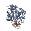
| ||||||||
|---|---|---|---|---|---|---|---|---|---|
| 1 |
| ||||||||
| Unit cell |
|
-Components
-DNA chain , 2 types, 2 molecules DE
| #1: DNA chain | Mass: 4635.010 Da / Num. of mol.: 1 / Source method: obtained synthetically |
|---|---|
| #2: DNA chain | Mass: 4398.854 Da / Num. of mol.: 1 / Source method: obtained synthetically |
-Protein , 1 types, 1 molecules A
| #3: Protein | Mass: 36394.176 Da / Num. of mol.: 1 / Fragment: CORE FRAGMENT (RESIDUES 12 TO 327) Source method: isolated from a genetically manipulated source Source: (gene. exp.) Homo sapiens (human) / Gene: ogg1 / Production host:  References: UniProt: O15527, Hydrolases; Glycosylases; Hydrolysing N-glycosyl compounds |
|---|
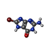
-Non-polymers , 3 types, 166 molecules 

| #4: Chemical | ChemComp-CA /  Mass: 40.078 Da / Num. of mol.: 1 / Source method: obtained synthetically / Formula: Ca Mass: 40.078 Da / Num. of mol.: 1 / Source method: obtained synthetically / Formula: Ca |
|---|---|
| #5: Chemical | ChemComp-BRG /  Mass: 230.022 Da / Num. of mol.: 1 / Source method: obtained synthetically / Formula: C5H4BrN5O Mass: 230.022 Da / Num. of mol.: 1 / Source method: obtained synthetically / Formula: C5H4BrN5O |
| #6: Water | ChemComp-HOH / Mass: 18.015 Da / Num. of mol.: 164 / Source method: isolated from a natural source / Formula: H2O |
-Details
| Has protein modification | Y |
|---|
-Experimental details
-Experiment
| Experiment | Method: X-RAY DIFFRACTION / Number of used crystals: 1 |
|---|
- Sample preparation
| Crystal | Density Matthews: 2.83 Å3/Da / Density % sol: 56.61 % | ||||||||||||||||||||||||||||||||||||||||||||||||
|---|---|---|---|---|---|---|---|---|---|---|---|---|---|---|---|---|---|---|---|---|---|---|---|---|---|---|---|---|---|---|---|---|---|---|---|---|---|---|---|---|---|---|---|---|---|---|---|---|---|
| Crystal grow | Temperature: 277 K / Method: vapor diffusion, hanging drop / pH: 6.3 Details: sodium cacodylate, calcium acetate, PEG 8000, pH 6.3, VAPOR DIFFUSION, HANGING DROP, temperature 277K | ||||||||||||||||||||||||||||||||||||||||||||||||
| Components of the solutions |
| ||||||||||||||||||||||||||||||||||||||||||||||||
| Crystal grow | *PLUS Temperature: 4 ℃ / pH: 7.4 / Details: Bruner, S.D., (2000) Nature, 403, 859. | ||||||||||||||||||||||||||||||||||||||||||||||||
| Components of the solutions | *PLUS
|
-Data collection
| Diffraction | Mean temperature: 100 K |
|---|---|
| Diffraction source | Source: SYNCHROTRON / Site: CHESS  / Beamline: A1 / Wavelength: 0.95 Å / Beamline: A1 / Wavelength: 0.95 Å |
| Detector | Type: ADSC QUANTUM 4 / Detector: CCD / Date: Dec 15, 2001 |
| Radiation | Protocol: SINGLE WAVELENGTH / Monochromatic (M) / Laue (L): M / Scattering type: x-ray |
| Radiation wavelength | Wavelength: 0.95 Å / Relative weight: 1 |
| Reflection | Resolution: 2.1→50 Å / Num. all: 32073 / Num. obs: 31837 / % possible obs: 99.3 % / Observed criterion σ(I): -3 / Redundancy: 5.4 % / Rmerge(I) obs: 0.101 / Net I/σ(I): 17.4 |
| Reflection shell | Resolution: 2.1→2.18 Å / Redundancy: 5.4 % / Rmerge(I) obs: 0.457 / Mean I/σ(I) obs: 4.9 / Num. unique all: 3096 / % possible all: 100 |
| Reflection | *PLUS Lowest resolution: 50 Å / Num. obs: 31356 |
| Reflection shell | *PLUS % possible obs: 100 % / Rmerge(I) obs: 0.46 |
- Processing
| Software |
| |||||||||||||||||||||||||
|---|---|---|---|---|---|---|---|---|---|---|---|---|---|---|---|---|---|---|---|---|---|---|---|---|---|---|
| Refinement | Method to determine structure: FOURIER SYNTHESIS Starting model: 1EBM Resolution: 2.1→50 Å / Cross valid method: THROUGHOUT / σ(F): 2 / Stereochemistry target values: Engh & Huber
| |||||||||||||||||||||||||
| Refine analyze |
| |||||||||||||||||||||||||
| Refinement step | Cycle: LAST / Resolution: 2.1→50 Å
| |||||||||||||||||||||||||
| Refine LS restraints |
| |||||||||||||||||||||||||
| LS refinement shell | Resolution: 2.1→2.18 Å
| |||||||||||||||||||||||||
| Refinement | *PLUS Lowest resolution: 50 Å / % reflection Rfree: 10 % / Rfactor Rfree: 0.257 / Rfactor Rwork: 0.236 | |||||||||||||||||||||||||
| Solvent computation | *PLUS | |||||||||||||||||||||||||
| Displacement parameters | *PLUS | |||||||||||||||||||||||||
| Refine LS restraints | *PLUS
|
