 Movie
Movie Controller

[English] 日本語
 Yorodumi
- PDB-3lau: Crystal Structure of Aurora2 kinase in complex with a GSK3beta in... -
+ Open data

- Basic information
| Entry | Database: PDB / ID: 3lau | ||||||
|---|---|---|---|---|---|---|---|
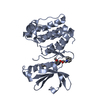
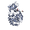
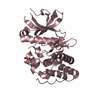
| Title | Crystal Structure of Aurora2 kinase in complex with a GSK3beta inhibitor | ||||||
 Components Components | Serine/threonine-protein kinase 6 | ||||||
 Keywords Keywords | TRANSFERASE / Kinase inhibitor complex / ATP-binding / Cell cycle / Cytoplasm / Cytoskeleton / Kinase / Nucleotide-binding / Phosphoprotein / Polymorphism / Serine/threonine-protein kinase / Ubl conjugation | ||||||
| Function / homology |  Function and homology information Function and homology informationInteraction between PHLDA1 and AURKA / regulation of centrosome cycle / axon hillock / spindle assembly involved in female meiosis I / cilium disassembly / spindle pole centrosome / histone H3S10 kinase activity / chromosome passenger complex / positive regulation of oocyte maturation / mitotic centrosome separation ...Interaction between PHLDA1 and AURKA / regulation of centrosome cycle / axon hillock / spindle assembly involved in female meiosis I / cilium disassembly / spindle pole centrosome / histone H3S10 kinase activity / chromosome passenger complex / positive regulation of oocyte maturation / mitotic centrosome separation / pronucleus / germinal vesicle / protein localization to centrosome / meiotic spindle / anterior/posterior axis specification / spindle organization / neuron projection extension / centrosome localization / positive regulation of mitochondrial fission / mitotic spindle pole / spindle midzone / SUMOylation of DNA replication proteins / negative regulation of protein binding / regulation of G2/M transition of mitotic cell cycle / positive regulation of mitotic nuclear division / protein serine/threonine/tyrosine kinase activity / positive regulation of mitotic cell cycle / TP53 Regulates Transcription of Genes Involved in G2 Cell Cycle Arrest / liver regeneration / molecular function activator activity / AURKA Activation by TPX2 / peptidyl-serine phosphorylation / regulation of signal transduction by p53 class mediator / mitotic spindle organization / regulation of cytokinesis / centriole / regulation of protein stability / APC/C:Cdh1 mediated degradation of Cdc20 and other APC/C:Cdh1 targeted proteins in late mitosis/early G1 / FBXL7 down-regulates AURKA during mitotic entry and in early mitosis / response to wounding / kinetochore / G2/M transition of mitotic cell cycle / spindle / spindle pole / mitotic spindle / protein autophosphorylation / Regulation of PLK1 Activity at G2/M Transition / mitotic cell cycle / positive regulation of proteasomal ubiquitin-dependent protein catabolic process / microtubule cytoskeleton / midbody / Regulation of TP53 Activity through Phosphorylation / basolateral plasma membrane / protein phosphorylation / microtubule / proteasome-mediated ubiquitin-dependent protein catabolic process / protein kinase activity / non-specific serine/threonine protein kinase / postsynaptic density / ciliary basal body / protein heterodimerization activity / negative regulation of gene expression / protein serine kinase activity / cell division / protein serine/threonine kinase activity / apoptotic process / ubiquitin protein ligase binding / centrosome / protein kinase binding / negative regulation of apoptotic process / perinuclear region of cytoplasm / glutamatergic synapse / nucleoplasm / ATP binding / nucleus / cytosol Similarity search - Function | ||||||
| Biological species |  Homo sapiens (human) Homo sapiens (human) | ||||||
| Method |  X-RAY DIFFRACTION / SYNCHROTRON / MOLECULAR REPLACEMENT / Resolution: 2.1 Å X-RAY DIFFRACTION / SYNCHROTRON / MOLECULAR REPLACEMENT / Resolution: 2.1 Å | ||||||
 Authors Authors | Maignan, S. / Guilloteau, J.P. / Pouzieux, S. | ||||||
 Citation Citation | Journal: Bioorg.Med.Chem.Lett. / Year: 2010 Title: Rational design of potent GSK3beta inhibitors with selectivity for Cdk1 and Cdk2. Authors: Lesuisse, D. / Dutruc-Rosset, G. / Tiraboschi, G. / Dreyer, M.K. / Maignan, S. / Chevalier, A. / Halley, F. / Bertrand, P. / Burgevin, M.C. / Quarteronet, D. / Rooney, T. | ||||||
| History |
|
- Structure visualization
| Structure viewer | Molecule: MolmilJmol/JSmol |
|---|
- Downloads & links
-Download
| PDBx/mmCIF format | 3lau.cif.gz | 69.3 KB | Display | PDBx/mmCIF format |
|---|---|---|---|---|
| PDB format | pdb3lau.ent.gz | 49.9 KB | Display | PDB format |
| PDBx/mmJSON format | 3lau.json.gz | Tree view | PDBx/mmJSON format | |
| Others |  Other downloads Other downloads |
-Validation report
| Arichive directory | https://data.pdbj.org/pub/pdb/validation_reports/la/3lauftp://data.pdbj.org/pub/pdb/validation_reports/la/3lau | HTTPS FTP |
|---|
-Related structure data
| Related structure data |  3lfnC  3lfqC  3lfsC 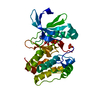 1mq4S S: Starting model for refinement C: citing same article ( |
|---|---|
| Similar structure data |


-Links
 PDBj
PDBj


- Assembly
| Deposited unit | 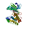
| ||||||||
|---|---|---|---|---|---|---|---|---|---|
| 1 |
| ||||||||
| Unit cell |
|
-Components
| #1: Protein | Mass: 33342.176 Da / Num. of mol.: 1 / Fragment: UNP residues 125-399 / Mutation: T288D Source method: isolated from a genetically manipulated source Source: (gene. exp.) Homo sapiens (human) / Gene: AIK, ARK1, AURA, AURKA, BTAK, STK15, STK6 / Production host:  References: UniProt: O14965, non-specific serine/threonine protein kinase |
|---|---|
| #2: Chemical | ChemComp-OFI /   Mass: 295.336 Da / Num. of mol.: 1 / Source method: obtained synthetically / Formula: C17H17N3O2 Mass: 295.336 Da / Num. of mol.: 1 / Source method: obtained synthetically / Formula: C17H17N3O2 |
| #3: Water | ChemComp-HOH /  Mass: 18.015 Da / Num. of mol.: 92 / Source method: isolated from a natural source / Formula: H2O Mass: 18.015 Da / Num. of mol.: 92 / Source method: isolated from a natural source / Formula: H2O |
-Experimental details
-Experiment
| Experiment | Method: X-RAY DIFFRACTION / Number of used crystals: 1 |
|---|
- Sample preparation
| Crystal | Density Matthews: 1.94 Å3/Da / Density % sol: 36.63 % |
|---|---|
| Crystal grow | Temperature: 298 K / Method: vapor diffusion, hanging drop / pH: 8.5 Details: PEG4000 26% - Tris 100mM pH 8.5 - DTT 5mM - MgCl2 1mM - NaAcetate 200mM, VAPOR DIFFUSION, HANGING DROP, temperature 298K |
-Data collection
| Diffraction | Mean temperature: 100 K |
|---|---|
| Diffraction source | Source: SYNCHROTRON / Site: ESRF  / Beamline: ID29 / Wavelength: 0.9 Å / Beamline: ID29 / Wavelength: 0.9 Å |
| Detector | Type: ccd / Detector: CCD / Date: Apr 1, 2002 |
| Radiation | Monochromator: Diamonds / Protocol: SINGLE WAVELENGTH / Monochromatic (M) / Laue (L): M / Scattering type: x-ray |
| Radiation wavelength | Wavelength: 0.9 Å / Relative weight: 1 |
| Reflection | Resolution: 2.1→30 Å / Num. obs: 13872 / % possible obs: 92.3 % / Observed criterion σ(F): 2 / Observed criterion σ(I): 2 / Redundancy: 3.5 % / Rsym value: 0.061 |
- Processing
| Software |
| ||||||||||||||||
|---|---|---|---|---|---|---|---|---|---|---|---|---|---|---|---|---|---|
| Refinement | Method to determine structure: MOLECULAR REPLACEMENT Starting model: PDB ENTRY 1MQ4 Resolution: 2.1→27.7 Å / Isotropic thermal model: Isotropic / Cross valid method: THROUGHOUT / σ(F): 2 / σ(I): 2 / Stereochemistry target values: Engh & Huber Details: CRYSTAL PACKING INTERACTIONS INDUCE A REARRANGEMENT OF SOME ACTIVATION LOOP RESIDUES (ARG255-HIS257) CLOSE TO THE BINDING SITE. THIS IS NOT OBSERVED IN OTHER CRYSTAL FORM AND CAN HAVE SOME ...Details: CRYSTAL PACKING INTERACTIONS INDUCE A REARRANGEMENT OF SOME ACTIVATION LOOP RESIDUES (ARG255-HIS257) CLOSE TO THE BINDING SITE. THIS IS NOT OBSERVED IN OTHER CRYSTAL FORM AND CAN HAVE SOME IMPACT ON LIGAND BINDING.
| ||||||||||||||||
| Displacement parameters | Biso mean: 32.2 Å2 | ||||||||||||||||
| Refinement step | Cycle: LAST / Resolution: 2.1→27.7 Å
|
