 Movie
Movie Controller

[English] 日本語
 Yorodumi
- PDB-4x3f: Crystal structure of the intracellular domain of the M. tuberculo... -
+ Open data

- Basic information
| Entry | Database: PDB / ID: 4x3f | ||||||
|---|---|---|---|---|---|---|---|
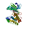
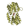
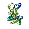
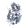
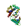
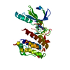
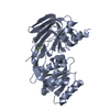
| Title | Crystal structure of the intracellular domain of the M. tuberculosis Ser/Thr kinase PknA | ||||||
 Components Components | (Serine/threonine-protein kinase PknA) x 3 | ||||||
 Keywords Keywords | TRANSFERASE / Kinase / autoinhibition / phosphorylation | ||||||
| Function / homology |  Function and homology information Function and homology informationpeptidyl-threonine autophosphorylation / negative regulation of lipid biosynthetic process / regulation of fatty acid metabolic process / negative regulation of fatty acid biosynthetic process / regulation of cell shape / non-specific serine/threonine protein kinase / protein serine kinase activity / protein serine/threonine kinase activity / regulation of DNA-templated transcription / extracellular region ...peptidyl-threonine autophosphorylation / negative regulation of lipid biosynthetic process / regulation of fatty acid metabolic process / negative regulation of fatty acid biosynthetic process / regulation of cell shape / non-specific serine/threonine protein kinase / protein serine kinase activity / protein serine/threonine kinase activity / regulation of DNA-templated transcription / extracellular region / ATP binding / plasma membrane / cytosol Similarity search - Function | ||||||
| Biological species |   Mycobacterium tuberculosis (bacteria) Mycobacterium tuberculosis (bacteria) | ||||||
| Method |  X-RAY DIFFRACTION / SYNCHROTRON / MOLECULAR REPLACEMENT / Resolution: 2.9 Å X-RAY DIFFRACTION / SYNCHROTRON / MOLECULAR REPLACEMENT / Resolution: 2.9 Å | ||||||
 Authors Authors | Wagner, T. / Wehenkel, A. / Alzari, P.M. / Bellinzoni, M. | ||||||
 Citation Citation | Journal: Proteins / Year: 2015 Title: The crystal structure of the catalytic domain of the ser/thr kinase PknA from M. tuberculosis shows an Src-like autoinhibited conformation. Authors: Wagner, T. / Alexandre, M. / Duran, R. / Barilone, N. / Wehenkel, A. / Alzari, P.M. / Bellinzoni, M. | ||||||
| History |
|
- Structure visualization
| Structure viewer | Molecule: MolmilJmol/JSmol |
|---|
- Downloads & links
-Download
| PDBx/mmCIF format | 4x3f.cif.gz | 321.1 KB | Display | PDBx/mmCIF format |
|---|---|---|---|---|
| PDB format | pdb4x3f.ent.gz | 261 KB | Display | PDB format |
| PDBx/mmJSON format | 4x3f.json.gz | Tree view | PDBx/mmJSON format | |
| Others |  Other downloads Other downloads |
-Validation report
| Arichive directory | https://data.pdbj.org/pub/pdb/validation_reports/x3/4x3fftp://data.pdbj.org/pub/pdb/validation_reports/x3/4x3f | HTTPS FTP |
|---|
-Related structure data
| Related structure data | 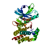 1o6yS S: Starting model for refinement |
|---|---|
| Similar structure data |


-Links
 PDBj
PDBj- Assembly
| Deposited unit | 
| |||||||||||||||||||||||||||||||||||||||||||||||||||||||||||||||||||||||||||||||||
|---|---|---|---|---|---|---|---|---|---|---|---|---|---|---|---|---|---|---|---|---|---|---|---|---|---|---|---|---|---|---|---|---|---|---|---|---|---|---|---|---|---|---|---|---|---|---|---|---|---|---|---|---|---|---|---|---|---|---|---|---|---|---|---|---|---|---|---|---|---|---|---|---|---|---|---|---|---|---|---|---|---|---|
| 1 | 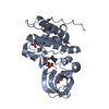
| |||||||||||||||||||||||||||||||||||||||||||||||||||||||||||||||||||||||||||||||||
| 2 | 
| |||||||||||||||||||||||||||||||||||||||||||||||||||||||||||||||||||||||||||||||||
| 3 | 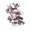
| |||||||||||||||||||||||||||||||||||||||||||||||||||||||||||||||||||||||||||||||||
| Unit cell |
| |||||||||||||||||||||||||||||||||||||||||||||||||||||||||||||||||||||||||||||||||
| Noncrystallographic symmetry (NCS) | NCS domain:
NCS domain segments: Component-ID: _ / End auth comp-ID: PRO / End label comp-ID: PRO / Refine code: _
NCS ensembles :
|
-Components
| #1: Protein | Mass: 36017.855 Da / Num. of mol.: 1 Source method: isolated from a genetically manipulated source Source: (gene. exp.) Mycobacterium tuberculosis (bacteria) / Strain: ATCC 25618 / H37Rv / Gene: pknA, Rv0015c, MTCY10H4.15c / Production host: References: UniProt: P9WI83, non-specific serine/threonine protein kinase |
|---|---|
| #2: Protein | Mass: 35937.875 Da / Num. of mol.: 1 Source method: isolated from a genetically manipulated source Source: (gene. exp.) Mycobacterium tuberculosis (bacteria) / Strain: ATCC 25618 / H37Rv / Gene: pknA, Rv0015c, MTCY10H4.15c / Production host: References: UniProt: P9WI83, non-specific serine/threonine protein kinase |
| #3: Protein | Mass: 35857.895 Da / Num. of mol.: 1 Source method: isolated from a genetically manipulated source Source: (gene. exp.) Mycobacterium tuberculosis (bacteria) / Strain: ATCC 25618 / H37Rv / Gene: pknA, Rv0015c, MTCY10H4.15c / Production host: References: UniProt: P9WI83, non-specific serine/threonine protein kinase |
| #4: Water | ChemComp-HOH /  Mass: 18.015 Da / Num. of mol.: 10 / Source method: isolated from a natural source / Formula: H2O Mass: 18.015 Da / Num. of mol.: 10 / Source method: isolated from a natural source / Formula: H2O |
| Has protein modification | Y |
-Experimental details
-Experiment
| Experiment | Method: X-RAY DIFFRACTION |
|---|
- Sample preparation
| Crystal | Density Matthews: 3.43 Å3/Da / Density % sol: 63.97 % |
|---|---|
| Crystal grow | Temperature: 291 K / Method: vapor diffusion, hanging drop / pH: 8 / Details: 4.3 M NaCl, 0.1 M Hepes-Na pH 8.0 |
-Data collection
| Diffraction | Mean temperature: 100 K |
|---|---|
| Diffraction source | Source: SYNCHROTRON / Site: ESRF  / Beamline: ID14-2 / Wavelength: 0.933 Å / Beamline: ID14-2 / Wavelength: 0.933 Å |
| Detector | Type: ADSC QUANTUM 210 / Detector: CCD / Date: Feb 9, 2008 |
| Radiation | Protocol: SINGLE WAVELENGTH / Monochromatic (M) / Laue (L): M / Scattering type: x-ray |
| Radiation wavelength | Wavelength: 0.933 Å / Relative weight: 1 |
| Reflection | Resolution: 2.9→43.6 Å / Num. obs: 31961 / % possible obs: 99.6 % / Redundancy: 3.6 % / Rmerge(I) obs: 0.107 / Net I/σ(I): 8.3 |
| Reflection shell | Resolution: 2.9→3.06 Å / Redundancy: 3.5 % / Rmerge(I) obs: 1.197 / Mean I/σ(I) obs: 1.1 / % possible all: 99.1 |
- Processing
| Software |
| ||||||||||||||||||||||||||||||||||||||||||||||||||||||||||||||||||||||||||||||||||||||||||||||||||||||||||||||||||||||||||||||||||||||||||||||||||||||||||||||||||||||||||||||||||||||
|---|---|---|---|---|---|---|---|---|---|---|---|---|---|---|---|---|---|---|---|---|---|---|---|---|---|---|---|---|---|---|---|---|---|---|---|---|---|---|---|---|---|---|---|---|---|---|---|---|---|---|---|---|---|---|---|---|---|---|---|---|---|---|---|---|---|---|---|---|---|---|---|---|---|---|---|---|---|---|---|---|---|---|---|---|---|---|---|---|---|---|---|---|---|---|---|---|---|---|---|---|---|---|---|---|---|---|---|---|---|---|---|---|---|---|---|---|---|---|---|---|---|---|---|---|---|---|---|---|---|---|---|---|---|---|---|---|---|---|---|---|---|---|---|---|---|---|---|---|---|---|---|---|---|---|---|---|---|---|---|---|---|---|---|---|---|---|---|---|---|---|---|---|---|---|---|---|---|---|---|---|---|---|---|
| Refinement | Method to determine structure: MOLECULAR REPLACEMENT Starting model: 1o6y Resolution: 2.9→43.56 Å / Cor.coef. Fo:Fc: 0.935 / Cor.coef. Fo:Fc free: 0.917 / SU B: 46.23 / SU ML: 0.365 / Cross valid method: THROUGHOUT / ESU R: 0.636 / ESU R Free: 0.32 / Stereochemistry target values: MAXIMUM LIKELIHOOD / Details: HYDROGENS HAVE BEEN ADDED IN THE RIDING POSITIONS
| ||||||||||||||||||||||||||||||||||||||||||||||||||||||||||||||||||||||||||||||||||||||||||||||||||||||||||||||||||||||||||||||||||||||||||||||||||||||||||||||||||||||||||||||||||||||
| Solvent computation | Ion probe radii: 0.8 Å / Shrinkage radii: 0.8 Å / VDW probe radii: 1.2 Å / Solvent model: MASK | ||||||||||||||||||||||||||||||||||||||||||||||||||||||||||||||||||||||||||||||||||||||||||||||||||||||||||||||||||||||||||||||||||||||||||||||||||||||||||||||||||||||||||||||||||||||
| Displacement parameters | Biso mean: 91.212 Å2
| ||||||||||||||||||||||||||||||||||||||||||||||||||||||||||||||||||||||||||||||||||||||||||||||||||||||||||||||||||||||||||||||||||||||||||||||||||||||||||||||||||||||||||||||||||||||
| Refinement step | Cycle: 1 / Resolution: 2.9→43.56 Å
| ||||||||||||||||||||||||||||||||||||||||||||||||||||||||||||||||||||||||||||||||||||||||||||||||||||||||||||||||||||||||||||||||||||||||||||||||||||||||||||||||||||||||||||||||||||||
| Refine LS restraints |
|