regulation of cytokinesis, actomyosin contractile ring assembly / centralspindlin complex / positive regulation of mitotic cytokinetic process / regulation of protein kinase activity / activation of protein kinase activity / regulation of attachment of spindle microtubules to kinetochore / bicellular tight junction assembly / activation of GTPase activity / regulation of small GTPase mediated signal transduction / RHOB GTPase cycle ...regulation of cytokinesis, actomyosin contractile ring assembly / centralspindlin complex / positive regulation of mitotic cytokinetic process / regulation of protein kinase activity / activation of protein kinase activity / regulation of attachment of spindle microtubules to kinetochore / bicellular tight junction assembly / activation of GTPase activity / regulation of small GTPase mediated signal transduction / RHOB GTPase cycle / NRAGE signals death through JNK / positive regulation of cytokinesis / cleavage furrow / mitotic cytokinesis / CDC42 GTPase cycle / positive regulation of GTPase activity / RHOA GTPase cycle / bicellular tight junction / RAC1 GTPase cycle / positive regulation of neuron differentiation / guanyl-nucleotide exchange factor activity / cellular response to calcium ion / GTPase activator activity / cellular response to ionizing radiation / protein homooligomerization / positive regulation of protein import into nucleus / small GTPase binding / cell morphogenesis / cellular response to hydrogen peroxide / cell-cell junction / mitotic spindle / nervous system development / protein transport / G alpha (12/13) signalling events / midbody / cell cortex / cell differentiation / positive regulation of canonical NF-kappaB signal transduction / intracellular signal transduction / nuclear body / positive regulation of apoptotic process / centrosome / protein homodimerization activity / nucleoplasm / nucleus / cytoplasm / cytosol Similarity search - Function
Resolution: 1.482→25 Å / Cor.coef. Fo:Fc: 0.959 / Cor.coef. Fo:Fc free: 0.95 / WRfactor Rfree: 0.192 / WRfactor Rwork: 0.173 / SU B: 1.098 / SU ML: 0.043 / Cross valid method: THROUGHOUT / σ(F): 0 / ESU R: 0.07 / ESU R Free: 0.071 / Stereochemistry target values: MAXIMUM LIKELIHOOD Details: HYDROGENS HAVE BEEN ADDED IN THE RIDING POSITIONS. U VALUES REFINED INDIVIDUALLY. ARP/WARP, COOT and the MOLPROBITY server were also used for refinement.
Rfactor
Num. reflection
% reflection
Selection details
Rfree
0.205
1570
4.879 %
thin shells (sftools)
Rwork
0.181
-
-
-
obs
0.182
32180
99.929 %
-
Solvent computation
Ion probe radii: 0.8 Å / Shrinkage radii: 0.8 Å / VDW probe radii: 1.4 Å / Solvent model: MASK BULK SOLVENT
Displacement parameters
Biso mean: 10.175 Å2
Baniso -1
Baniso -2
Baniso -3
1-
-0.296 Å2
0 Å2
0 Å2
2-
-
-0.163 Å2
0 Å2
3-
-
-
0.459 Å2
Refinement step
Cycle: LAST / Resolution: 1.482→25 Å
Protein
Nucleic acid
Ligand
Solvent
Total
Num. atoms
1463
0
3
119
1585
Refine LS restraints
Refine-ID
Type
Dev ideal
Dev ideal target
Number
X-RAY DIFFRACTION
r_bond_refined_d
0.016
0.022
1538
X-RAY DIFFRACTION
r_bond_other_d
0.001
0.02
1032
X-RAY DIFFRACTION
r_angle_refined_deg
1.551
1.967
2096
X-RAY DIFFRACTION
r_angle_other_deg
0.885
3
2537
X-RAY DIFFRACTION
r_dihedral_angle_1_deg
6.028
5
197
X-RAY DIFFRACTION
r_dihedral_angle_2_deg
30.905
25.616
73
X-RAY DIFFRACTION
r_dihedral_angle_3_deg
12.018
15
261
X-RAY DIFFRACTION
r_dihedral_angle_4_deg
13.13
15
4
X-RAY DIFFRACTION
r_chiral_restr
0.087
0.2
224
X-RAY DIFFRACTION
r_gen_planes_refined
0.007
0.021
1727
X-RAY DIFFRACTION
r_gen_planes_other
0.001
0.02
301
X-RAY DIFFRACTION
r_mcbond_it
0.941
1.5
941
X-RAY DIFFRACTION
r_mcbond_other
0.26
1.5
378
X-RAY DIFFRACTION
r_mcangle_it
1.665
2
1526
X-RAY DIFFRACTION
r_scbond_it
2.604
3
597
X-RAY DIFFRACTION
r_scangle_it
4.148
4.5
563
LS refinement shell
Refine-ID: X-RAY DIFFRACTION / Total num. of bins used: 20
Resolution (Å)
Rfactor Rfree
Num. reflection Rfree
Rfactor Rwork
Num. reflection Rwork
Num. reflection all
% reflection obs (%)
1.482-1.52
0.293
136
0.234
2169
2324
99.182
1.52-1.562
0.208
116
0.212
2159
2276
99.956
1.562-1.607
0.245
101
0.213
2125
2226
100
1.607-1.656
0.223
106
0.188
2022
2129
99.953
1.656-1.71
0.178
107
0.174
2000
2107
100
1.71-1.77
0.202
115
0.179
1921
2037
99.951
1.77-1.836
0.228
104
0.175
1865
1969
100
1.836-1.911
0.188
112
0.173
1754
1866
100
1.911-1.995
0.189
115
0.171
1713
1828
100
1.995-2.092
0.242
36
0.174
1712
1748
100
2.092-2.204
0.205
92
0.171
1558
1650
100
2.204-2.337
0.222
56
0.171
1533
1589
100
2.337-2.496
0.203
54
0.178
1425
1479
100
2.496-2.694
0.233
75
0.184
1308
1383
100
2.694-2.948
0.237
55
0.21
1233
1288
100
2.948-3.29
0.239
52
0.185
1131
1183
100
3.29-3.788
0.16
52
0.166
987
1039
100
3.788-4.613
0.131
29
0.144
877
906
100
4.613-6.416
0.156
31
0.189
688
719
100
6.416-25
0.247
26
0.199
430
457
99.781
+
About Yorodumi
-
News
-
Feb 9, 2022. New format data for meta-information of EMDB entries
New format data for meta-information of EMDB entries
Version 3 of the EMDB header file is now the official format.
The previous official version 1.9 will be removed from the archive.
In the structure databanks used in Yorodumi, some data are registered as the other names, "COVID-19 virus" and "2019-nCoV". Here are the details of the virus and the list of structure data.
Jan 31, 2019. EMDB accession codes are about to change! (news from PDBe EMDB page)
EMDB accession codes are about to change! (news from PDBe EMDB page)
The allocation of 4 digits for EMDB accession codes will soon come to an end. Whilst these codes will remain in use, new EMDB accession codes will include an additional digit and will expand incrementally as the available range of codes is exhausted. The current 4-digit format prefixed with “EMD-” (i.e. EMD-XXXX) will advance to a 5-digit format (i.e. EMD-XXXXX), and so on. It is currently estimated that the 4-digit codes will be depleted around Spring 2019, at which point the 5-digit format will come into force.
The EM Navigator/Yorodumi systems omit the EMD- prefix.
Related info.:Q: What is EMD? / ID/Accession-code notation in Yorodumi/EM Navigator
Yorodumi is a browser for structure data from EMDB, PDB, SASBDB, etc.
This page is also the successor to EM Navigator detail page, and also detail information page/front-end page for Omokage search.
The word "yorodu" (or yorozu) is an old Japanese word meaning "ten thousand". "mi" (miru) is to see.
Related info.:EMDB / PDB / SASBDB / Comparison of 3 databanks / Yorodumi Search / Aug 31, 2016. New EM Navigator & Yorodumi / Yorodumi Papers / Jmol/JSmol / Function and homology information / Changes in new EM Navigator and Yorodumi
 Movie
Movie Controller

 Yorodumi
 Open data
 Basic information
 Components
 Keywords
 Function and homology information
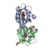
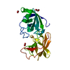
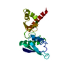
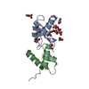
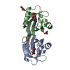
 Homo sapiens (human)
 X-RAY DIFFRACTION /
  Authors
 Citation
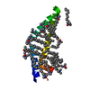
 Structure visualization
 Downloads & links
 Other downloads
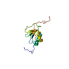


 PDBj
PDBj


 Assembly
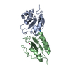


 Num. of mol.: 3 / Source method: obtained synthetically
 Mass: 18.015 Da / Num. of mol.: 119 / Source method: isolated from a natural source / Formula: H2O
 Sample preparation
 / Beamline: 19-ID / Wavelength: 0.97934 Å
 Processing