Mass: 18.015 Da / Num. of mol.: 65 / Source method: isolated from a natural source / Formula: H2O
Has protein modification
Y
Nonpolymer details
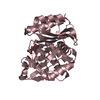
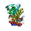
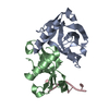
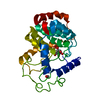
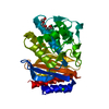
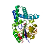
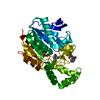
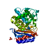
ALTHOUGH THE REAL SPACE R VALUE FOR SOME OF THE LDA RESIDUES ARE HIGH, THESE LDAO DETERGENT ...ALTHOUGH THE REAL SPACE R VALUE FOR SOME OF THE LDA RESIDUES ARE HIGH, THESE LDAO DETERGENT MOLECULES (RESIDUE NAME IS LDA) ARE PART OF A MICELLE-TYPE STRUCTURE, AND THE OBSERVED DENSITY FOR THESE LIGANDS IS COMPLETELY CONSISTENT WITH THE PACKING REQUIREMENTS OF THE NEIGHBORING LDA MOLECULES.
-
Experimental details
-
Experiment
Experiment
Method: X-RAY DIFFRACTION / Number of used crystals: 1
-
Sample preparation
Crystal
Density Matthews: 3.12 Å3/Da / Density % sol: 60.56 %
Crystal grow
Temperature: 300 K / Method: vapor diffusion / pH: 6.5 Details: 1.6 M trisodium citrate, pH 6.5, VAPOR DIFFUSION, temperature 300K
Resolution: 1.9→1.93 Å / Redundancy: 5.1 % / Rmerge(I) obs: 0.443 / Mean I/σ(I) obs: 3.2 / % possible all: 98.1
-
Processing
Software
Name
Version
Classification
NB
REFMAC
5.5.0102
refinement
PDB_EXTRACT
3.1
dataextraction
HKL-3000
datacollection
HKL-3000
datareduction
HKL-3000
datascaling
SHELXS
phasing
Refinement
Method to determine structure: SAD / Resolution: 1.9→10 Å / Cor.coef. Fo:Fc: 0.939 / Cor.coef. Fo:Fc free: 0.913 / Occupancy max: 1 / Occupancy min: 0.5 / SU B: 8.515 / SU ML: 0.118 / Cross valid method: THROUGHOUT / σ(F): 0 / ESU R Free: 0.176 / Stereochemistry target values: MAXIMUM LIKELIHOOD Details: HYDROGENS HAVE BEEN ADDED IN THE RIDING POSITIONS U VALUES: RESIDUAL ONLY
Rfactor
Num. reflection
% reflection
Selection details
Rfree
0.28
478
4.8 %
RANDOM
Rwork
0.23
-
-
-
obs
0.232
9889
99.3 %
-
all
-
9889
-
-
Solvent computation
Ion probe radii: 0.8 Å / Shrinkage radii: 0.8 Å / VDW probe radii: 1.4 Å / Solvent model: MASK
Displacement parameters
Biso mean: 41.21 Å2
Baniso -1
Baniso -2
Baniso -3
1-
1.05 Å2
0.52 Å2
0 Å2
2-
-
1.05 Å2
0 Å2
3-
-
-
-1.57 Å2
Refinement step
Cycle: LAST / Resolution: 1.9→10 Å
Protein
Nucleic acid
Ligand
Solvent
Total
Num. atoms
606
0
320
65
991
Refine LS restraints
Refine-ID
Type
Dev ideal
Dev ideal target
Number
X-RAY DIFFRACTION
r_bond_refined_d
0.017
0.021
917
X-RAY DIFFRACTION
r_bond_other_d
X-RAY DIFFRACTION
r_angle_refined_deg
1.9
2.301
1179
X-RAY DIFFRACTION
r_angle_other_deg
X-RAY DIFFRACTION
r_dihedral_angle_1_deg
4.576
5
79
X-RAY DIFFRACTION
r_dihedral_angle_2_deg
40.434
27.727
22
X-RAY DIFFRACTION
r_dihedral_angle_3_deg
14.674
15
116
X-RAY DIFFRACTION
r_dihedral_angle_4_deg
10.914
15
1
X-RAY DIFFRACTION
r_chiral_restr
0.112
0.2
101
X-RAY DIFFRACTION
r_gen_planes_refined
0.008
0.021
439
X-RAY DIFFRACTION
r_gen_planes_other
X-RAY DIFFRACTION
r_nbd_refined
X-RAY DIFFRACTION
r_nbd_other
X-RAY DIFFRACTION
r_nbtor_refined
X-RAY DIFFRACTION
r_nbtor_other
X-RAY DIFFRACTION
r_xyhbond_nbd_refined
X-RAY DIFFRACTION
r_xyhbond_nbd_other
X-RAY DIFFRACTION
r_metal_ion_refined
X-RAY DIFFRACTION
r_metal_ion_other
X-RAY DIFFRACTION
r_symmetry_vdw_refined
X-RAY DIFFRACTION
r_symmetry_vdw_other
X-RAY DIFFRACTION
r_symmetry_hbond_refined
X-RAY DIFFRACTION
r_symmetry_hbond_other
X-RAY DIFFRACTION
r_symmetry_metal_ion_refined
X-RAY DIFFRACTION
r_symmetry_metal_ion_other
X-RAY DIFFRACTION
r_mcbond_it
1.196
1.5
401
X-RAY DIFFRACTION
r_mcbond_other
X-RAY DIFFRACTION
r_mcangle_it
2.19
2
653
X-RAY DIFFRACTION
r_scbond_it
2.825
3
516
X-RAY DIFFRACTION
r_scangle_it
4.55
4.5
526
X-RAY DIFFRACTION
r_rigid_bond_restr
X-RAY DIFFRACTION
r_sphericity_free
X-RAY DIFFRACTION
r_sphericity_bonded
LS refinement shell
Resolution: 1.9→1.95 Å / Total num. of bins used: 20
Rfactor
Num. reflection
% reflection
Rfree
0.274
32
-
Rwork
0.208
630
-
obs
-
-
98.66 %
Refinement TLS params.
Method: refined / Refine-ID: X-RAY DIFFRACTION
ID
L11 (°2)
L12 (°2)
L13 (°2)
L22 (°2)
L23 (°2)
L33 (°2)
S11 (Å °)
S12 (Å °)
S13 (Å °)
S21 (Å °)
S22 (Å °)
S23 (Å °)
S31 (Å °)
S32 (Å °)
S33 (Å °)
T11 (Å2)
T12 (Å2)
T13 (Å2)
T22 (Å2)
T23 (Å2)
T33 (Å2)
Origin x (Å)
Origin y (Å)
Origin z (Å)
1
1.1858
-0.2517
-0.1908
3.4612
0.8737
1.381
0.0239
0.0514
0.0521
0.0016
0.1416
-0.4036
0.007
0.0121
-0.1656
0.0322
-0.0092
0.0042
0.0918
-0.048
0.0751
29.558
-4.286
-16.28
2
0.5204
0.3499
1.3122
1.1137
1.0582
3.3582
0.0852
-0.0733
-0.0298
0.0086
-0.0142
-0.0046
0.1719
-0.1482
-0.071
0.0279
-0.0311
-0.001
0.1063
-0.0328
0.0511
20.227
13.5
0.31
3
0.8933
0.4957
0.8833
1.2505
1.2001
2.4564
-0.1062
-0.2654
-0.023
-0.0576
-0.1162
0.3045
0.3517
-0.1002
0.2224
0.2401
0.0466
0.0261
0.1346
0.0083
0.1099
22.584
-0.148
-4.168
Refinement TLS group
ID
Refine-ID
Refine TLS-ID
Auth asym-ID
Auth seq-ID
1
X-RAY DIFFRACTION
1
A
1 - 17
2
X-RAY DIFFRACTION
1
A
68 - 80
3
X-RAY DIFFRACTION
2
A
18 - 67
4
X-RAY DIFFRACTION
3
A
101
5
X-RAY DIFFRACTION
3
A
102
6
X-RAY DIFFRACTION
3
A
103
7
X-RAY DIFFRACTION
3
A
104
8
X-RAY DIFFRACTION
3
A
105
9
X-RAY DIFFRACTION
3
A
106
10
X-RAY DIFFRACTION
3
A
107
11
X-RAY DIFFRACTION
3
A
108
12
X-RAY DIFFRACTION
3
A
109
13
X-RAY DIFFRACTION
3
A
110
14
X-RAY DIFFRACTION
3
A
111
15
X-RAY DIFFRACTION
3
A
112
16
X-RAY DIFFRACTION
3
A
113
17
X-RAY DIFFRACTION
3
A
114
18
X-RAY DIFFRACTION
3
A
115
19
X-RAY DIFFRACTION
3
A
116
20
X-RAY DIFFRACTION
3
A
117
21
X-RAY DIFFRACTION
3
A
118
22
X-RAY DIFFRACTION
3
A
119
23
X-RAY DIFFRACTION
3
A
120
+
About Yorodumi
-
News
-
Feb 9, 2022. New format data for meta-information of EMDB entries
New format data for meta-information of EMDB entries
Version 3 of the EMDB header file is now the official format.
The previous official version 1.9 will be removed from the archive.
In the structure databanks used in Yorodumi, some data are registered as the other names, "COVID-19 virus" and "2019-nCoV". Here are the details of the virus and the list of structure data.
Jan 31, 2019. EMDB accession codes are about to change! (news from PDBe EMDB page)
EMDB accession codes are about to change! (news from PDBe EMDB page)
The allocation of 4 digits for EMDB accession codes will soon come to an end. Whilst these codes will remain in use, new EMDB accession codes will include an additional digit and will expand incrementally as the available range of codes is exhausted. The current 4-digit format prefixed with “EMD-” (i.e. EMD-XXXX) will advance to a 5-digit format (i.e. EMD-XXXXX), and so on. It is currently estimated that the 4-digit codes will be depleted around Spring 2019, at which point the 5-digit format will come into force.
The EM Navigator/Yorodumi systems omit the EMD- prefix.
Related info.:Q: What is EMD? / ID/Accession-code notation in Yorodumi/EM Navigator
Yorodumi is a browser for structure data from EMDB, PDB, SASBDB, etc.
This page is also the successor to EM Navigator detail page, and also detail information page/front-end page for Omokage search.
The word "yorodu" (or yorozu) is an old Japanese word meaning "ten thousand". "mi" (miru) is to see.
Related info.:EMDB / PDB / SASBDB / Comparison of 3 databanks / Yorodumi Search / Aug 31, 2016. New EM Navigator & Yorodumi / Yorodumi Papers / Jmol/JSmol / Function and homology information / Changes in new EM Navigator and Yorodumi
 Movie
Movie Controller

 Yorodumi
 Open data
 Basic information
 Components
 Keywords
 Function and homology information
 Homo sapiens (human)
 X-RAY DIFFRACTION /
  Authors
 Citation
 Structure visualization
 Downloads & links
 Other downloads
 PDBj
PDBj


 Assembly


 Mass: 229.402 Da / Num. of mol.: 20 / Source method: obtained synthetically / Formula: C14H31NO / Comment: LDAO, detergent*YM
 Mass: 18.015 Da / Num. of mol.: 65 / Source method: isolated from a natural source / Formula: H2O
 Sample preparation
 Processing