 Movie
Movie Controller

[English] 日本語
 Yorodumi
- PDB-2jc7: The crystal structure of the carbapenemase OXA-24 reveals new ins... -
+ Open data

- Basic information
| Entry | Database: PDB / ID: 2jc7 | ||||||
|---|---|---|---|---|---|---|---|
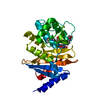
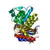
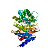
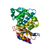
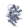
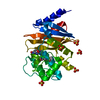
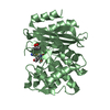
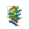
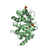
| Title | The crystal structure of the carbapenemase OXA-24 reveals new insights into the mechanism of carbapenem-hydrolysis | ||||||
 Components Components | BETA-LACTAMASE OXA-24 | ||||||
 Keywords Keywords | HYDROLASE / PLASMID / B-LACTAMASES / ENZYME MECHANISM / CARBAPENEM RESISTANCE | ||||||
| Function / homology |  Function and homology information Function and homology information | ||||||
| Biological species |  ACINETOBACTER BAUMANNII (bacteria) ACINETOBACTER BAUMANNII (bacteria) | ||||||
| Method |  X-RAY DIFFRACTION / SYNCHROTRON / MOLECULAR REPLACEMENT / Resolution: 2.5 Å X-RAY DIFFRACTION / SYNCHROTRON / MOLECULAR REPLACEMENT / Resolution: 2.5 Å | ||||||
 Authors Authors | Santillana, E. / Romero, A. | ||||||
 Citation Citation | Journal: Proc.Natl.Acad.Sci.USA / Year: 2007 Title: Crystal Structure of the Carbapenemase Oxa-24 Reveals Insights Into the Mechanism of Carbapenem Hydrolysis. Authors: Santillana, E. / Beceiro, A. / Bou, G. / Romero, A. | ||||||
| History |
|
- Structure visualization
| Structure viewer | Molecule: MolmilJmol/JSmol |
|---|
- Downloads & links
-Download
| PDBx/mmCIF format | 2jc7.cif.gz | 63.1 KB | Display | PDBx/mmCIF format |
|---|---|---|---|---|
| PDB format | pdb2jc7.ent.gz | 45.5 KB | Display | PDB format |
| PDBx/mmJSON format | 2jc7.json.gz | Tree view | PDBx/mmJSON format | |
| Others |  Other downloads Other downloads |
-Validation report
| Arichive directory | https://data.pdbj.org/pub/pdb/validation_reports/jc/2jc7ftp://data.pdbj.org/pub/pdb/validation_reports/jc/2jc7 | HTTPS FTP |
|---|
-Related structure data
| Related structure data | 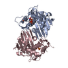 1k57S S: Starting model for refinement |
|---|---|
| Similar structure data |

-Links
 PDBj
PDBj


- Assembly
| Deposited unit | 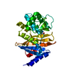
| ||||||||
|---|---|---|---|---|---|---|---|---|---|
| 1 |
| ||||||||
| Unit cell |
|
-Components
| #1: Protein | Mass: 27543.658 Da / Num. of mol.: 1 / Fragment: RESIDUES 32-275 Source method: isolated from a genetically manipulated source Source: (gene. exp.) ACINETOBACTER BAUMANNII (bacteria) / Plasmid: PGEX-6P-1 / Production host: |
|---|---|
| #2: Chemical | ChemComp-SO4 /   Mass: 96.063 Da / Num. of mol.: 1 / Source method: obtained synthetically / Formula: SO4 Mass: 96.063 Da / Num. of mol.: 1 / Source method: obtained synthetically / Formula: SO4 |
| #3: Water | ChemComp-HOH /  Mass: 18.015 Da / Num. of mol.: 121 / Source method: isolated from a natural source / Formula: H2O Mass: 18.015 Da / Num. of mol.: 121 / Source method: isolated from a natural source / Formula: H2O |
| Sequence details | RESIDUES 1 TO 21 COMPRISED THE SIGNAL PEPTIDE AND ARE NOT PRESENT IN THE RECOMBINANT PROTEIN ...RESIDUES 1 TO 21 COMPRISED THE SIGNAL PEPTIDE AND ARE NOT PRESENT IN THE RECOMBINAN |
-Experimental details
-Experiment
| Experiment | Method: X-RAY DIFFRACTION / Number of used crystals: 1 |
|---|
- Sample preparation
| Crystal | Density Matthews: 4.09 Å3/Da / Density % sol: 68.75 % |
|---|---|
| Crystal grow | Details: 0.1 M BIS-TRIS PH 6.5, 2 M AMMONIUM SULFATE |
-Data collection
| Diffraction | Mean temperature: 110 K |
|---|---|
| Diffraction source | Source: SYNCHROTRON / Site: ESRF  / Beamline: BM16 / Wavelength: 0.979 / Beamline: BM16 / Wavelength: 0.979 |
| Detector | Type: MARRESEARCH / Detector: CCD |
| Radiation | Protocol: SINGLE WAVELENGTH / Monochromatic (M) / Laue (L): M / Scattering type: x-ray |
| Radiation wavelength | Wavelength: 0.979 Å / Relative weight: 1 |
| Reflection | Resolution: 2.5→55 Å / Num. obs: 16630 / % possible obs: 99.4 % / Observed criterion σ(I): 0 / Redundancy: 13.9 % / Biso Wilson estimate: 36.4 Å2 / Rmerge(I) obs: 0.1 / Net I/σ(I): 5.4 |
| Reflection shell | Resolution: 2.48→2.62 Å / Redundancy: 13.4 % / Rmerge(I) obs: 0.25 / Mean I/σ(I) obs: 2.9 / % possible all: 96.3 |
- Processing
| Software |
| ||||||||||||||||||||||||||||||||||||||||||||||||||||||||||||||||||||||||||||||||
|---|---|---|---|---|---|---|---|---|---|---|---|---|---|---|---|---|---|---|---|---|---|---|---|---|---|---|---|---|---|---|---|---|---|---|---|---|---|---|---|---|---|---|---|---|---|---|---|---|---|---|---|---|---|---|---|---|---|---|---|---|---|---|---|---|---|---|---|---|---|---|---|---|---|---|---|---|---|---|---|---|---|
| Refinement | Method to determine structure: MOLECULAR REPLACEMENT Starting model: PDB ENTRY 1K57 Resolution: 2.5→15 Å / Rfactor Rfree error: 0.007 / Data cutoff high absF: 1439570.96 / Data cutoff low absF: 0 / Isotropic thermal model: RESTRAINED / Cross valid method: THROUGHOUT / σ(F): 0
| ||||||||||||||||||||||||||||||||||||||||||||||||||||||||||||||||||||||||||||||||
| Solvent computation | Solvent model: FLAT MODEL / Bsol: 35.4546 Å2 / ksol: 0.384349 e/Å3 | ||||||||||||||||||||||||||||||||||||||||||||||||||||||||||||||||||||||||||||||||
| Displacement parameters | Biso mean: 32.1 Å2
| ||||||||||||||||||||||||||||||||||||||||||||||||||||||||||||||||||||||||||||||||
| Refine analyze |
| ||||||||||||||||||||||||||||||||||||||||||||||||||||||||||||||||||||||||||||||||
| Refinement step | Cycle: LAST / Resolution: 2.5→15 Å
| ||||||||||||||||||||||||||||||||||||||||||||||||||||||||||||||||||||||||||||||||
| Refine LS restraints |
| ||||||||||||||||||||||||||||||||||||||||||||||||||||||||||||||||||||||||||||||||
| LS refinement shell | Resolution: 2.5→2.66 Å / Rfactor Rfree error: 0.021 / Total num. of bins used: 6
| ||||||||||||||||||||||||||||||||||||||||||||||||||||||||||||||||||||||||||||||||
| Xplor file |
|
