 Movie
Movie Controller


+ Open data

- Basic information
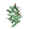
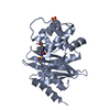
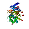
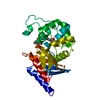
| Entry | Database: PDB / ID: 3g4p | ||||||
|---|---|---|---|---|---|---|---|
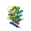
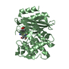
| Title | OXA-24 beta-lactamase at pH 7.5 | ||||||
 Components Components | Beta-lactamase OXA-24 | ||||||
 Keywords Keywords | HYDROLASE / B-LACTAMASES / ENZYME MECHANISM / CARBAPENEM / RESISTANCE | ||||||
| Function / homology |  Function and homology information Function and homology information | ||||||
| Biological species |  Acinetobacter baumannii (bacteria) Acinetobacter baumannii (bacteria) | ||||||
| Method |  X-RAY DIFFRACTION / SYNCHROTRON / MOLECULAR REPLACEMENT / Resolution: 1.97 Å X-RAY DIFFRACTION / SYNCHROTRON / MOLECULAR REPLACEMENT / Resolution: 1.97 Å | ||||||
 Authors Authors | Santillana, E. / Romero, A. | ||||||
 Citation Citation | Journal: J.Am.Chem.Soc. / Year: 2010 Title: Design, synthesis, and crystal structures of 6-alkylidene-2'-substituted penicillanic acid sulfones as potent inhibitors of Acinetobacter baumannii OXA-24 carbapenemase Authors: Bou, G. / Santillana, E. / Sheri, A. / Beceiro, A. / Sampson, J.M. / Kalp, M. / Bethel, C.R. / Distler, A.M. / Drawz, S.M. / Pagadala, S.R. / van den Akker, F. / Bonomo, R.A. / Romero, A. / Buynak, J.D. #1: Journal: Proc.Natl.Acad.Sci.USA / Year: 2007Title: Crystal structure of the carbapenemase OXA-24 reveals insights into the mechanism of carbapenem hydrolysis Authors: Santillana, E. / Beceiro, A. / Bou, G. / Romero, A. | ||||||
| History |
|
- Structure visualization
| Structure viewer | Molecule: MolmilJmol/JSmol |
|---|
- Downloads & links
-Download
| PDBx/mmCIF format | 3g4p.cif.gz | 65.5 KB | Display | PDBx/mmCIF format |
|---|---|---|---|---|
| PDB format | pdb3g4p.ent.gz | 47.5 KB | Display | PDB format |
| PDBx/mmJSON format | 3g4p.json.gz | Tree view | PDBx/mmJSON format | |
| Others |  Other downloads Other downloads |
-Validation report
| Arichive directory | https://data.pdbj.org/pub/pdb/validation_reports/g4/3g4pftp://data.pdbj.org/pub/pdb/validation_reports/g4/3g4p | HTTPS FTP |
|---|
-Related structure data
| Related structure data |  3fv7C  3fyzC  3fzcC  3mbzC  2jc7S S: Starting model for refinement C: citing same article ( |
|---|---|
| Similar structure data |


-Links
 PDBj
PDBj


- Assembly
| Deposited unit | 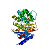
| ||||||||
|---|---|---|---|---|---|---|---|---|---|
| 1 |
| ||||||||
| Unit cell |
| ||||||||
| Components on special symmetry positions |
|
-Components
| #1: Protein | Mass: 27586.660 Da / Num. of mol.: 1 / Fragment: UNP RESIDUES 32-275 Source method: isolated from a genetically manipulated source Source: (gene. exp.) Acinetobacter baumannii (bacteria) / Gene: oxa-24 / Plasmid: PGEX-6P-1 / Production host: |
|---|---|
| #2: Water | ChemComp-HOH /  Mass: 18.015 Da / Num. of mol.: 167 / Source method: isolated from a natural source / Formula: H2O Mass: 18.015 Da / Num. of mol.: 167 / Source method: isolated from a natural source / Formula: H2O |
-Experimental details
-Experiment
| Experiment | Method: X-RAY DIFFRACTION / Number of used crystals: 1 |
|---|
- Sample preparation
| Crystal | Density Matthews: 3.98 Å3/Da / Density % sol: 69.13 % |
|---|---|
| Crystal grow | Temperature: 293 K / Method: vapor diffusion, sitting drop / pH: 7.5 Details: 0.1M SODIUM ACETATE, 28% PEG 2000 MME, 0.1M HEPES (PH 7.5), VAPOR DIFFUSION, SITTING DROP, temperature 293K |
-Data collection
| Diffraction | Mean temperature: 100 K |
|---|---|
| Diffraction source | Source: SYNCHROTRON / Site: ESRF  / Beamline: ID14-2 / Wavelength: 0.933 Å / Beamline: ID14-2 / Wavelength: 0.933 Å |
| Detector | Type: ADSC QUANTUM 4 / Detector: CCD / Date: Jun 14, 2008 |
| Radiation | Monochromator: SINGLE SILICON (111) / Protocol: SINGLE WAVELENGTH / Monochromatic (M) / Laue (L): M / Scattering type: x-ray |
| Radiation wavelength | Wavelength: 0.933 Å / Relative weight: 1 |
| Reflection | Resolution: 1.97→31.59 Å / Num. all: 32135 / Num. obs: 32064 / % possible obs: 99.8 % / Observed criterion σ(F): 0 / Observed criterion σ(I): 0 / Redundancy: 11.7 % / Biso Wilson estimate: 24.04 Å2 / Rmerge(I) obs: 0.086 / Net I/σ(I): 7.2 |
| Reflection shell | Resolution: 1.97→2.08 Å / Redundancy: 11.2 % / Rmerge(I) obs: 0.509 / Mean I/σ(I) obs: 1.5 / Num. unique all: 4548 / % possible all: 99.5 |
- Processing
| Software |
| ||||||||||||||||||||||||||||||||||||||||||||||||||||||||||||||||||||||||||||||||||||||||||
|---|---|---|---|---|---|---|---|---|---|---|---|---|---|---|---|---|---|---|---|---|---|---|---|---|---|---|---|---|---|---|---|---|---|---|---|---|---|---|---|---|---|---|---|---|---|---|---|---|---|---|---|---|---|---|---|---|---|---|---|---|---|---|---|---|---|---|---|---|---|---|---|---|---|---|---|---|---|---|---|---|---|---|---|---|---|---|---|---|---|---|---|
| Refinement | Method to determine structure: MOLECULAR REPLACEMENT Starting model: PDB ENTRY 2jc7 Resolution: 1.97→30.95 Å / Cor.coef. Fo:Fc: 0.957 / Cor.coef. Fo:Fc free: 0.933 / SU B: 3.702 / SU ML: 0.1 / Cross valid method: THROUGHOUT / σ(F): 0 / ESU R: 0.122 / ESU R Free: 0.129 / Stereochemistry target values: MAXIMUM LIKELIHOOD / Details: HYDROGENS HAVE BEEN ADDED IN THE RIDING POSITIONS
| ||||||||||||||||||||||||||||||||||||||||||||||||||||||||||||||||||||||||||||||||||||||||||
| Solvent computation | Ion probe radii: 0.8 Å / Shrinkage radii: 0.8 Å / VDW probe radii: 1.2 Å / Solvent model: MASK | ||||||||||||||||||||||||||||||||||||||||||||||||||||||||||||||||||||||||||||||||||||||||||
| Displacement parameters | Biso mean: 30 Å2
| ||||||||||||||||||||||||||||||||||||||||||||||||||||||||||||||||||||||||||||||||||||||||||
| Refinement step | Cycle: LAST / Resolution: 1.97→30.95 Å
| ||||||||||||||||||||||||||||||||||||||||||||||||||||||||||||||||||||||||||||||||||||||||||
| Refine LS restraints |
| ||||||||||||||||||||||||||||||||||||||||||||||||||||||||||||||||||||||||||||||||||||||||||
| LS refinement shell | Resolution: 1.97→2.021 Å / Total num. of bins used: 20
|
