 Movie
Movie Controller

[English] 日本語
 Yorodumi
- PDB-3l3o: Staphylococcal Complement Inhibitor (SCIN) in complex with Human ... -
+ Open data

- Basic information
| Entry | Database: PDB / ID: 3l3o | ||||||
|---|---|---|---|---|---|---|---|
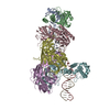
| Title | Staphylococcal Complement Inhibitor (SCIN) in complex with Human Complement Component C3c | ||||||
 Components Components |
| ||||||
 Keywords Keywords | IMMUNE SYSTEM / Complement alternate pathway / Complement pathway / Converstase / Immune response / Inflammatory response / Innate immunity / Secreted / Virulence / Immune evasion | ||||||
| Function / homology |  Function and homology information Function and homology informationC5L2 anaphylatoxin chemotactic receptor binding / oviduct epithelium development / regulation of triglyceride biosynthetic process / positive regulation of activation of membrane attack complex / vertebrate eye-specific patterning / positive regulation of apoptotic cell clearance / Alternative complement activation / complement-mediated synapse pruning / positive regulation of type IIa hypersensitivity / Activation of C3 and C5 ...C5L2 anaphylatoxin chemotactic receptor binding / oviduct epithelium development / regulation of triglyceride biosynthetic process / positive regulation of activation of membrane attack complex / vertebrate eye-specific patterning / positive regulation of apoptotic cell clearance / Alternative complement activation / complement-mediated synapse pruning / positive regulation of type IIa hypersensitivity / Activation of C3 and C5 / positive regulation of lipid storage / complement activation, GZMK pathway / positive regulation of phagocytosis, engulfment / positive regulation of G protein-coupled receptor signaling pathway / complement-dependent cytotoxicity / positive regulation of D-glucose transmembrane transport / complement receptor mediated signaling pathway / complement activation, alternative pathway / complement activation / endopeptidase inhibitor activity / neuron remodeling / amyloid-beta clearance / B cell activation / complement activation, classical pathway / positive regulation of vascular endothelial growth factor production / Purinergic signaling in leishmaniasis infection / Regulation of Complement cascade / Peptide ligand-binding receptors / response to bacterium / Post-translational protein phosphorylation / fatty acid metabolic process / positive regulation of protein phosphorylation / positive regulation of receptor-mediated endocytosis / Regulation of Insulin-like Growth Factor (IGF) transport and uptake by Insulin-like Growth Factor Binding Proteins (IGFBPs) / Immunoregulatory interactions between a Lymphoid and a non-Lymphoid cell / positive regulation of angiogenesis / azurophil granule lumen / secretory granule lumen / blood microparticle / G alpha (i) signalling events / immune response / endoplasmic reticulum lumen / G protein-coupled receptor signaling pathway / inflammatory response / receptor ligand activity / signaling receptor binding / Neutrophil degranulation / cell surface / signal transduction / protein-containing complex / : / extracellular exosome / extracellular region / plasma membrane Similarity search - Function | ||||||
| Biological species |   Staphylococcus aureus (bacteria) Staphylococcus aureus (bacteria) Homo sapiens (human) Homo sapiens (human) | ||||||
| Method |  X-RAY DIFFRACTION / SYNCHROTRON / MOLECULAR REPLACEMENT / molecular replacement / Resolution: 3.405 Å X-RAY DIFFRACTION / SYNCHROTRON / MOLECULAR REPLACEMENT / molecular replacement / Resolution: 3.405 Å | ||||||
 Authors Authors | Geisbrecht, B.V. / Garcia, B.G. | ||||||
 Citation Citation | Journal: J.Mol.Biol. / Year: 2010 Title: Molecular Basis for Complement Recognition and Inhibition Determined by Crystallographic Studies of the Staphylococcal Complement Inhibitor (SCIN) Bound to C3c and C3b. Authors: Garcia, B.L. / Ramyar, K.X. / Tzekou, A. / Ricklin, D. / McWhorter, W.J. / Lambris, J.D. / Geisbrecht, B.V. | ||||||
| History |
|
- Structure visualization
| Structure viewer | Molecule: MolmilJmol/JSmol |
|---|
- Downloads & links
-Download
| PDBx/mmCIF format | 3l3o.cif.gz | 489 KB | Display | PDBx/mmCIF format |
|---|---|---|---|---|
| PDB format | pdb3l3o.ent.gz | 389.6 KB | Display | PDB format |
| PDBx/mmJSON format | 3l3o.json.gz | Tree view | PDBx/mmJSON format | |
| Others |  Other downloads Other downloads |
-Validation report
| Arichive directory | https://data.pdbj.org/pub/pdb/validation_reports/l3/3l3oftp://data.pdbj.org/pub/pdb/validation_reports/l3/3l3o | HTTPS FTP |
|---|
-Related structure data
| Related structure data |  3l5nC  3nmsC  3ohxC 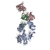 2a74S  2qffS C: citing same article ( S: Starting model for refinement |
|---|---|
| Similar structure data |


-Links
 PDBj
PDBj


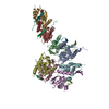
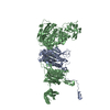
- Assembly
| Deposited unit | 
| ||||||||
|---|---|---|---|---|---|---|---|---|---|
| 1 | 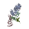
| ||||||||
| 2 | 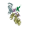
| ||||||||
| Unit cell |
|
-Components
| #1: Protein | Mass: 71393.320 Da / Num. of mol.: 2 / Fragment: residues 23-667 / Source method: isolated from a natural source / Source: (natural) Homo sapiens (human) / References: UniProt: P01024#2: Protein | Mass: 23621.244 Da / Num. of mol.: 2 / Fragment: residues 749-954 / Source method: isolated from a natural source / Source: (natural) Homo sapiens (human) / References: UniProt: P01024#3: Protein | Mass: 10064.370 Da / Num. of mol.: 2 Source method: isolated from a genetically manipulated source Source: (gene. exp.) Staphylococcus aureus (bacteria) / Strain: Mu50 / ATCC 700699 / Gene: SAV1942, scn / Plasmid: pt7HMT / Production host: #4: Protein | Mass: 39537.418 Da / Num. of mol.: 2 / Fragment: residues 1321-1663 / Source method: isolated from a natural source / Source: (natural) Homo sapiens (human) / References: UniProt: P01024#5: Sugar |   Type: D-saccharide, beta linking / Mass: 221.208 Da / Num. of mol.: 2 Type: D-saccharide, beta linking / Mass: 221.208 Da / Num. of mol.: 2Source method: isolated from a genetically manipulated source Formula: C8H15NO6 Has protein modification | Y | |
|---|
-Experimental details
-Experiment
| Experiment | Method: X-RAY DIFFRACTION / Number of used crystals: 1 |
|---|
- Sample preparation
| Crystal | Density Matthews: 3 Å3/Da / Density % sol: 58.98 % |
|---|---|
| Crystal grow | Temperature: 277 K / Method: vapor diffusion, hanging drop / pH: 7.5 Details: Protein solution: 5mg/ml, 0.1M HEPES, 10% PEG 6000, 5% 2-Methyl-2,4-pentanediol, pH 7.5, VAPOR DIFFUSION, HANGING DROP, temperature 277K |
-Data collection
| Diffraction | Mean temperature: 93 K | |||||||||||||||||||||||||||||||||||||||||||||||||||||||||||||||||||||||||||||
|---|---|---|---|---|---|---|---|---|---|---|---|---|---|---|---|---|---|---|---|---|---|---|---|---|---|---|---|---|---|---|---|---|---|---|---|---|---|---|---|---|---|---|---|---|---|---|---|---|---|---|---|---|---|---|---|---|---|---|---|---|---|---|---|---|---|---|---|---|---|---|---|---|---|---|---|---|---|---|
| Diffraction source | Source: SYNCHROTRON / Site: APS  / Beamline: 22-BM / Wavelength: 1 Å / Beamline: 22-BM / Wavelength: 1 Å | |||||||||||||||||||||||||||||||||||||||||||||||||||||||||||||||||||||||||||||
| Detector | Type: MARMOSAIC 225 mm CCD / Detector: CCD / Date: Jun 12, 2009 / Details: mirrors | |||||||||||||||||||||||||||||||||||||||||||||||||||||||||||||||||||||||||||||
| Radiation | Monochromator: Si(220) / Protocol: SINGLE WAVELENGTH / Monochromatic (M) / Laue (L): M / Scattering type: x-ray | |||||||||||||||||||||||||||||||||||||||||||||||||||||||||||||||||||||||||||||
| Radiation wavelength | Wavelength: 1 Å / Relative weight: 1 | |||||||||||||||||||||||||||||||||||||||||||||||||||||||||||||||||||||||||||||
| Reflection | Redundancy: 3.4 % / Av σ(I) over netI: 6.5 / Number: 152670 / Rmerge(I) obs: 0.161 / Χ2: 1.11 / D res high: 3.4 Å / D res low: 50 Å / Num. obs: 45397 / % possible obs: 97.5 | |||||||||||||||||||||||||||||||||||||||||||||||||||||||||||||||||||||||||||||
| Diffraction reflection shell |
| |||||||||||||||||||||||||||||||||||||||||||||||||||||||||||||||||||||||||||||
| Reflection | Resolution: 3.4→50 Å / Num. all: 48213 / Num. obs: 45397 / % possible obs: 97.5 % / Observed criterion σ(F): 2.38 / Observed criterion σ(I): 2.38 / Redundancy: 3.4 % / Biso Wilson estimate: 49.8 Å2 / Rmerge(I) obs: 0.161 / Net I/σ(I): 5 | |||||||||||||||||||||||||||||||||||||||||||||||||||||||||||||||||||||||||||||
| Reflection shell | Resolution: 3.4→3.52 Å / Redundancy: 2.8 % / Rmerge(I) obs: 0.497 / Mean I/σ(I) obs: 2.38 / % possible all: 93.1 |
-Phasing
| Phasing | Method: molecular replacement | |||||||||
|---|---|---|---|---|---|---|---|---|---|---|
| Phasing MR | Rfactor: 41.99 / Model details: Phaser MODE: MR_AUTO
|
- Processing
| Software |
| ||||||||||||||||||||||||||||||||||||||||||||||||||||||||||||||||||||||||||||||||||||||||||||||||||
|---|---|---|---|---|---|---|---|---|---|---|---|---|---|---|---|---|---|---|---|---|---|---|---|---|---|---|---|---|---|---|---|---|---|---|---|---|---|---|---|---|---|---|---|---|---|---|---|---|---|---|---|---|---|---|---|---|---|---|---|---|---|---|---|---|---|---|---|---|---|---|---|---|---|---|---|---|---|---|---|---|---|---|---|---|---|---|---|---|---|---|---|---|---|---|---|---|---|---|---|
| Refinement | Method to determine structure: MOLECULAR REPLACEMENT Starting model: 2A74,2QFF Resolution: 3.405→34.522 Å / Occupancy max: 1 / Occupancy min: 1 / SU ML: 2.38 / σ(F): 0.21 / Phase error: 27.44 / Stereochemistry target values: ML
| ||||||||||||||||||||||||||||||||||||||||||||||||||||||||||||||||||||||||||||||||||||||||||||||||||
| Solvent computation | Shrinkage radii: 0.9 Å / VDW probe radii: 1.11 Å / Solvent model: FLAT BULK SOLVENT MODEL / Bsol: 40.458 Å2 / ksol: 0.256 e/Å3 | ||||||||||||||||||||||||||||||||||||||||||||||||||||||||||||||||||||||||||||||||||||||||||||||||||
| Displacement parameters | Biso mean: 108.06 Å2
| ||||||||||||||||||||||||||||||||||||||||||||||||||||||||||||||||||||||||||||||||||||||||||||||||||
| Refinement step | Cycle: LAST / Resolution: 3.405→34.522 Å
| ||||||||||||||||||||||||||||||||||||||||||||||||||||||||||||||||||||||||||||||||||||||||||||||||||
| Refine LS restraints |
| ||||||||||||||||||||||||||||||||||||||||||||||||||||||||||||||||||||||||||||||||||||||||||||||||||
| LS refinement shell |
|
