 Movie
Movie Controller

[English] 日本語
 Yorodumi
- PDB-3kxb: Structural characterization of H3K56Q nucleosomes and nucleosomal... -
+ Open data

- Basic information
| Entry | Database: PDB / ID: 3kxb | ||||||
|---|---|---|---|---|---|---|---|
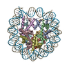
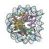
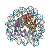
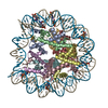
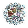
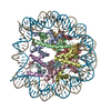
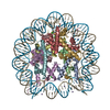
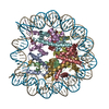
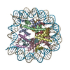
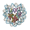
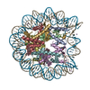
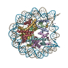
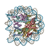
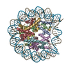
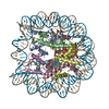
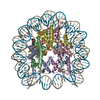
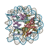
| Title | Structural characterization of H3K56Q nucleosomes and nucleosomal arrays | ||||||
 Components Components |
| ||||||
 Keywords Keywords | Transcription/DNA / Nucleosome / Transcription / Transcription-DNA complex | ||||||
| Function / homology |  Function and homology information Function and homology informationstructural constituent of chromatin / nucleosome / nucleosome assembly / heterochromatin formation / protein heterodimerization activity / DNA binding / nucleoplasm / nucleus Similarity search - Function | ||||||
| Biological species | |||||||
| Method |  X-RAY DIFFRACTION / MOLECULAR REPLACEMENT / Resolution: 3.2 Å X-RAY DIFFRACTION / MOLECULAR REPLACEMENT / Resolution: 3.2 Å | ||||||
 Authors Authors | Clark, N.J. / Lilyestrom, W.G. | ||||||
 Citation Citation | Journal: Biochim.Biophys.Acta / Year: 2010 Title: Structural characterization of H3K56Q nucleosomes and nucleosomal arrays. Authors: Watanabe, S. / Resch, M. / Lilyestrom, W. / Clark, N. / Hansen, J.C. / Peterson, C. / Luger, K. | ||||||
| History |
|
- Structure visualization
| Structure viewer | Molecule: MolmilJmol/JSmol |
|---|
- Downloads & links
-Download
| PDBx/mmCIF format | 3kxb.cif.gz | 315.1 KB | Display | PDBx/mmCIF format |
|---|---|---|---|---|
| PDB format | pdb3kxb.ent.gz | 241.8 KB | Display | PDB format |
| PDBx/mmJSON format | 3kxb.json.gz | Tree view | PDBx/mmJSON format | |
| Others |  Other downloads Other downloads |
-Validation report
| Arichive directory | https://data.pdbj.org/pub/pdb/validation_reports/kx/3kxbftp://data.pdbj.org/pub/pdb/validation_reports/kx/3kxb | HTTPS FTP |
|---|
-Related structure data
| Related structure data |  3kwqC 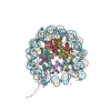 1aoiS S: Starting model for refinement C: citing same article ( |
|---|---|
| Similar structure data |


-Links
 PDBj
PDBj


- Assembly
| Deposited unit | 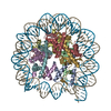
| ||||||||
|---|---|---|---|---|---|---|---|---|---|
| 1 |
| ||||||||
| Unit cell |
| ||||||||
| Details | A single nucleosome is in the asymmetric unit. One nucleosome is composed of 2 chains each of the four histones (H2A, H2B, H3 and H4) in an octamer as well as 146 base pairs of double stranded DNA. No symmetry operations are required to build the biological unit. |
-Components
-Protein , 4 types, 8 molecules AEBFCGDH
| #1: Protein | Mass: 15303.863 Da / Num. of mol.: 2 / Mutation: K57E, G103A Source method: isolated from a genetically manipulated source Source: (gene. exp.)  #2: Protein | Mass: 11263.231 Da / Num. of mol.: 2 Source method: isolated from a genetically manipulated source Source: (gene. exp.) #3: Protein | Mass: 13978.241 Da / Num. of mol.: 2 Source method: isolated from a genetically manipulated source Source: (gene. exp.) #4: Protein | Mass: 13524.752 Da / Num. of mol.: 2 / Mutation: S33T Source method: isolated from a genetically manipulated source Source: (gene. exp.) |
|---|
-DNA chain / Non-polymers , 2 types, 117 molecules IJ
| #5: DNA chain | Mass: 45054.844 Da / Num. of mol.: 2 / Source method: obtained synthetically #6: Water | ChemComp-HOH / | Mass: 18.015 Da / Num. of mol.: 115 / Source method: isolated from a natural source / Formula: H2O |
|---|
-Experimental details
-Experiment
| Experiment | Method: X-RAY DIFFRACTION / Number of used crystals: 1 |
|---|
- Sample preparation
| Crystal | Density Matthews: 2.65 Å3/Da / Density % sol: 53.52 % |
|---|---|
| Crystal grow | Temperature: 293 K / Method: vapor diffusion / pH: 6 Details: Crystals were grown by vapor diffusion in 8 20 days at 20 C using a droplet containing 4.0 mg ml−1 core particle 50 mM KCl, 70 75 mM MnCl , and 20 mM potassium cacodylate, pH 6.0, ...Details: Crystals were grown by vapor diffusion in 8 20 days at 20 C using a droplet containing 4.0 mg ml−1 core particle 50 mM KCl, 70 75 mM MnCl , and 20 mM potassium cacodylate, pH 6.0, surrounded by silicon oil DC200 (110mPa s; Fluka) and equilibrated against 40 46 mM MnCl2, 35 40 mM KCl and 20 mM potassium cacodylate, pH 6.0, VAPOR DIFFUSION, temperature 293K |
-Data collection
| Diffraction | Mean temperature: 100 K |
|---|---|
| Diffraction source | Source: ROTATING ANODE / Type: RIGAKU RUH3R / Wavelength: 1.54 Å |
| Detector | Type: RIGAKU RAXIS IV++ / Detector: IMAGE PLATE / Date: Oct 17, 2007 / Details: Rigaku Varimax HR |
| Radiation | Protocol: SINGLE WAVELENGTH / Monochromatic (M) / Laue (L): M / Scattering type: x-ray |
| Radiation wavelength | Wavelength: 1.54 Å / Relative weight: 1 |
| Reflection | Resolution: 3.2→20 Å / Num. all: 35254 / Num. obs: 35254 / % possible obs: 100 % / Observed criterion σ(F): 2 / Observed criterion σ(I): 2 / Redundancy: 1.3 % / Biso Wilson estimate: 79.7 Å2 / Rmerge(I) obs: 0.055 / Rsym value: 0.055 / Net I/σ(I): 19.53 |
| Reflection shell | Highest resolution: 3.2 Å / % possible all: 78 |
- Processing
| Software |
| |||||||||||||||||||||||||
|---|---|---|---|---|---|---|---|---|---|---|---|---|---|---|---|---|---|---|---|---|---|---|---|---|---|---|
| Refinement | Method to determine structure: MOLECULAR REPLACEMENT Starting model: PDBID 1AOI Resolution: 3.2→20 Å / Cross valid method: THROUGHOUT / σ(F): 2 / σ(I): 2 / Stereochemistry target values: Engh & Huber
| |||||||||||||||||||||||||
| Displacement parameters | Biso mean: 79.7 Å2 | |||||||||||||||||||||||||
| Refinement step | Cycle: LAST / Resolution: 3.2→20 Å
|
