 Movie
Movie Controller

[English] 日本語
 Yorodumi
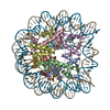
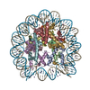
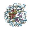
- PDB-3kwq: Structural characterization of H3K56Q nucleosomes and nucleosomal... -
+ Open data

- Basic information
| Entry | Database: PDB / ID: 3kwq | ||||||
|---|---|---|---|---|---|---|---|
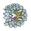
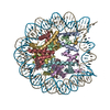
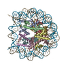
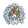
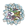
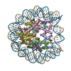
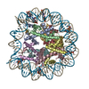
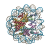
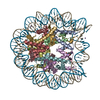
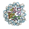
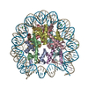
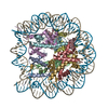
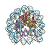
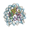
| Title | Structural characterization of H3K56Q nucleosomes and nucleosomal arrays | ||||||
 Components Components |
| ||||||
 Keywords Keywords | Structural Protein/DNA / Nucleosome Transcription K56 mutation / Acetylation / Chromosomal protein / DNA-binding / Methylation / Nucleosome core / Nucleus / Phosphoprotein / Isopeptide bond / Ubl conjugation / Structural Protein-DNA complex | ||||||
| Function / homology |  Function and homology information Function and homology informationstructural constituent of chromatin / nucleosome / heterochromatin formation / nucleosome assembly / protein heterodimerization activity / DNA binding / nucleoplasm / nucleus Similarity search - Function | ||||||
| Biological species | |||||||
| Method |  X-RAY DIFFRACTION / SYNCHROTRON / MOLECULAR REPLACEMENT / Resolution: 3.5 Å X-RAY DIFFRACTION / SYNCHROTRON / MOLECULAR REPLACEMENT / Resolution: 3.5 Å | ||||||
 Authors Authors | Lilyestrom, W.G. / Clark, N. | ||||||
 Citation Citation | Journal: Biochim.Biophys.Acta Title: Structural characterization of H3K56Q nucleosomes and nucleosomal arrays. Authors: Watanabe, S. / Resch, M. / Lilyestrom, W. / Clark, N. / Hansen, J.C. / Peterson, C. / Luger, K. | ||||||
| History |
|
- Structure visualization
| Structure viewer | Molecule: MolmilJmol/JSmol |
|---|
- Downloads & links
-Download
| PDBx/mmCIF format | 3kwq.cif.gz | 313.1 KB | Display | PDBx/mmCIF format |
|---|---|---|---|---|
| PDB format | pdb3kwq.ent.gz | 242.6 KB | Display | PDB format |
| PDBx/mmJSON format | 3kwq.json.gz | Tree view | PDBx/mmJSON format | |
| Others |  Other downloads Other downloads |
-Validation report
| Summary document | 3kwq_validation.pdf.gz | 495.9 KB | Display | wwPDB validaton report |
|---|---|---|---|---|
| Full document | 3kwq_full_validation.pdf.gz | 535.7 KB | Display | |
| Data in XML | 3kwq_validation.xml.gz | 39.3 KB | Display | |
| Data in CIF | 3kwq_validation.cif.gz | 55.7 KB | Display | |
| Arichive directory | https://data.pdbj.org/pub/pdb/validation_reports/kw/3kwqftp://data.pdbj.org/pub/pdb/validation_reports/kw/3kwq | HTTPS FTP |
-Related structure data
| Related structure data | 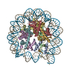 3kxbC 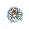 1aoiS S: Starting model for refinement C: citing same article ( |
|---|---|
| Similar structure data |


-Links
 PDBj
PDBj


- Assembly
| Deposited unit | 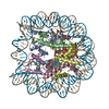
| ||||||||
|---|---|---|---|---|---|---|---|---|---|
| 1 |
| ||||||||
| Unit cell |
|
-Components
-Protein , 4 types, 8 molecules AEBFCGDH
| #1: Protein | Mass: 11502.369 Da / Num. of mol.: 2 / Fragment: UNP residues 39-136 / Mutation: K57E Source method: isolated from a genetically manipulated source Source: (gene. exp.)  #2: Protein | Mass: 9409.056 Da / Num. of mol.: 2 / Fragment: UNP residues 21-103 Source method: isolated from a genetically manipulated source Source: (gene. exp.) #3: Protein | Mass: 11724.677 Da / Num. of mol.: 2 / Fragment: UNP residues 15-121 Source method: isolated from a genetically manipulated source Source: (gene. exp.) #4: Protein | Mass: 10376.928 Da / Num. of mol.: 2 / Fragment: UNP residues 34-126 Source method: isolated from a genetically manipulated source Source: (gene. exp.) |
|---|
-DNA chain / Non-polymers , 2 types, 25 molecules IJ
| #5: DNA chain | Mass: 45054.844 Da / Num. of mol.: 2 / Source method: obtained synthetically #6: Water | ChemComp-HOH / | Mass: 18.015 Da / Num. of mol.: 23 / Source method: isolated from a natural source / Formula: H2O |
|---|
-Details
| Sequence details | THE AUTHORS CLONE CONTAIN THE DEVIATION G102A FROM THE UNP SEQUENCE |
|---|
-Experimental details
-Experiment
| Experiment | Method: X-RAY DIFFRACTION / Number of used crystals: 1 |
|---|
- Sample preparation
| Crystal | Density Matthews: 2.96 Å3/Da / Density % sol: 58.5 % |
|---|
-Data collection
| Diffraction | Mean temperature: 100 K |
|---|---|
| Diffraction source | Source: SYNCHROTRON / Site: ALS  / Beamline: 4.2.2 / Wavelength: 1 Å / Beamline: 4.2.2 / Wavelength: 1 Å |
| Detector | Type: NOIR-1 / Detector: CCD / Date: May 5, 2006 |
| Radiation | Scattering type: x-ray |
| Radiation wavelength | Wavelength: 1 Å / Relative weight: 1 |
| Reflection | Resolution: 3.5→20 Å / Num. all: 42705 / Num. obs: 42705 / % possible obs: 100 % / Observed criterion σ(F): 2 / Observed criterion σ(I): 2 / Redundancy: 7.23 % / Biso Wilson estimate: 87.5 Å2 / Rmerge(I) obs: 0.109 / Rsym value: 0.109 / Net I/σ(I): 6 |
- Processing
| Software |
| |||||||||||||||||||||||||
|---|---|---|---|---|---|---|---|---|---|---|---|---|---|---|---|---|---|---|---|---|---|---|---|---|---|---|
| Refinement | Method to determine structure: MOLECULAR REPLACEMENT Starting model: PDBID 1AOI Resolution: 3.5→20 Å / Cross valid method: THROUGHOUT / σ(F): 2 / σ(I): 2 / Stereochemistry target values: Engh & Huber
| |||||||||||||||||||||||||
| Displacement parameters | Biso mean: 87.5 Å2 | |||||||||||||||||||||||||
| Refinement step | Cycle: LAST / Resolution: 3.5→20 Å
|
