 Movie
Movie Controller

[English] 日本語
 Yorodumi
- PDB-3ko1: Cystal structure of thermosome from Acidianus tengchongensis strain S5 -
+ Open data

- Basic information
| Entry | Database: PDB / ID: 3ko1 | ||||||
|---|---|---|---|---|---|---|---|
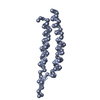
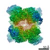
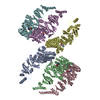
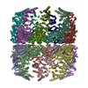
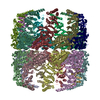
| Title | Cystal structure of thermosome from Acidianus tengchongensis strain S5 | ||||||
 Components Components | Chaperonin | ||||||
 Keywords Keywords | CHAPERONE / 9-fold symmetry / double ring / ATP hydrolase / Nucleotide-binding | ||||||
| Function / homology |  Function and homology information Function and homology informationchaperonin ATPase / ATP-dependent protein folding chaperone / : / ATP hydrolysis activity / protein-containing complex / ATP binding / metal ion binding / identical protein binding / cytoplasm Similarity search - Function | ||||||
| Biological species |  Acidianus tengchongensis (archaea) Acidianus tengchongensis (archaea) | ||||||
| Method |  X-RAY DIFFRACTION / SYNCHROTRON / MOLECULAR REPLACEMENT / Resolution: 3.7 Å X-RAY DIFFRACTION / SYNCHROTRON / MOLECULAR REPLACEMENT / Resolution: 3.7 Å | ||||||
 Authors Authors | Huo, Y. / Zhang, K. / Hu, Z. / Wang, L. / Zhai, Y. / Zhou, Q. / Lander, G. / He, Y. / Zhu, J. / Xu, W. ...Huo, Y. / Zhang, K. / Hu, Z. / Wang, L. / Zhai, Y. / Zhou, Q. / Lander, G. / He, Y. / Zhu, J. / Xu, W. / Dong, Z. / Sun, F. | ||||||
 Citation Citation | Journal: Structure / Year: 2010 Title: Crystal structure of group II chaperonin in the open state. Authors: Yanwu Huo / Zhongjun Hu / Kai Zhang / Li Wang / Yujia Zhai / Qiangjun Zhou / Gabe Lander / Jiang Zhu / Yongzhi He / Xiaoyun Pang / Wei Xu / Mark Bartlam / Zhiyang Dong / Fei Sun /  Abstract: Thermosomes are group II chaperonins responsible for protein refolding in an ATP-dependent manner. Little is known regarding the conformational changes of thermosomes during their functional cycle ...Thermosomes are group II chaperonins responsible for protein refolding in an ATP-dependent manner. Little is known regarding the conformational changes of thermosomes during their functional cycle due to a lack of high-resolution structure in the open state. Here, we report the first complete crystal structure of thermosome (rATcpnβ) in the open state from Acidianus tengchongensis. There is a ∼30° rotation of the apical and lid domains compared with the previous closed structure. Besides, the structure reveals a conspicuous hydrophobic patch in the lid domain, and residues locating in this patch are conserved across species. Both the closed and open forms of rATcpnβ were also reconstructed by electron microscopy (EM). Structural fitting revealed the detailed conformational change from the open to the closed state. Structural comparison as well as protease K digestion indicated only ATP binding without hydrolysis does not induce chamber closure of thermosome. | ||||||
| History |
|
- Structure visualization
| Structure viewer | Molecule: MolmilJmol/JSmol |
|---|
- Downloads & links
-Download
| PDBx/mmCIF format | 3ko1.cif.gz | 748.5 KB | Display | PDBx/mmCIF format |
|---|---|---|---|---|
| PDB format | pdb3ko1.ent.gz | 627.2 KB | Display | PDB format |
| PDBx/mmJSON format | 3ko1.json.gz | Tree view | PDBx/mmJSON format | |
| Others |  Other downloads Other downloads |
-Validation report
| Arichive directory | https://data.pdbj.org/pub/pdb/validation_reports/ko/3ko1ftp://data.pdbj.org/pub/pdb/validation_reports/ko/3ko1 | HTTPS FTP |
|---|
-Related structure data
| Related structure data | 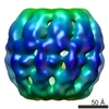 5154C 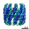 5157C 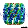 5159C 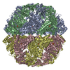 1q3rS S: Starting model for refinement C: citing same article ( |
|---|---|
| Similar structure data |

-Links
 PDBj
PDBj
- Assembly
| Deposited unit | 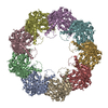
| ||||||||||||||||||||||||||||||
|---|---|---|---|---|---|---|---|---|---|---|---|---|---|---|---|---|---|---|---|---|---|---|---|---|---|---|---|---|---|---|---|
| 1 | 
| ||||||||||||||||||||||||||||||
| Unit cell |
| ||||||||||||||||||||||||||||||
| Noncrystallographic symmetry (NCS) | NCS domain:
|
-Components
| #1: Protein | Mass: 59738.344 Da / Num. of mol.: 9 Source method: isolated from a genetically manipulated source Source: (gene. exp.) Acidianus tengchongensis (archaea) / Strain: S5 / Plasmid: pET23b / Production host:  #2: Chemical | ChemComp-ADP /   Mass: 427.201 Da / Num. of mol.: 9 / Source method: obtained synthetically / Formula: C10H15N5O10P2 / Comment: ADP, energy-carrying molecule*YM Mass: 427.201 Da / Num. of mol.: 9 / Source method: obtained synthetically / Formula: C10H15N5O10P2 / Comment: ADP, energy-carrying molecule*YM |
|---|
-Experimental details
-Experiment
| Experiment | Method: X-RAY DIFFRACTION / Number of used crystals: 1 |
|---|
- Sample preparation
| Crystal | Density Matthews: 3.43 Å3/Da / Density % sol: 64.14 % |
|---|---|
| Crystal grow | Temperature: 289 K / Method: vapor diffusion, hanging drop / pH: 8.5 Details: 0.1M Tris-Hcl, 1.15M Li2SO4, pH 8.5, VAPOR DIFFUSION, HANGING DROP, temperature 289K |
-Data collection
| Diffraction | Mean temperature: 100 K |
|---|---|
| Diffraction source | Source: SYNCHROTRON / Site: Photon Factory  / Beamline: BL-17A / Wavelength: 0.9 Å / Beamline: BL-17A / Wavelength: 0.9 Å |
| Detector | Type: ADSC QUANTUM 270 / Detector: CCD / Date: Sep 10, 2009 |
| Radiation | Monochromator: Si(111) double crystal / Protocol: SINGLE WAVELENGTH / Monochromatic (M) / Laue (L): M / Scattering type: x-ray |
| Radiation wavelength | Wavelength: 0.9 Å / Relative weight: 1 |
| Reflection | Resolution: 3.7→50 Å / Num. obs: 76576 / % possible obs: 99.5 % / Observed criterion σ(I): 0 / Redundancy: 3.2 % / Biso Wilson estimate: 58 Å2 / Rmerge(I) obs: 0.117 / Net I/σ(I): 7.4 |
| Reflection shell | Resolution: 3.7→3.9 Å / Redundancy: 3.3 % / Rmerge(I) obs: 0.476 / Mean I/σ(I) obs: 2.3 / Num. unique all: 3429 / % possible all: 99.4 |
- Processing
| Software |
| |||||||||||||||||||||
|---|---|---|---|---|---|---|---|---|---|---|---|---|---|---|---|---|---|---|---|---|---|---|
| Refinement | Method to determine structure: MOLECULAR REPLACEMENT Starting model: 1Q3R Resolution: 3.7→48.39 Å / SU B: 47.548 / SU ML: 0.649 / Cross valid method: THROUGHOUT / σ(F): 0 / ESU R Free: 0.814 / Stereochemistry target values: MAXIMUM LIKELIHOOD / Details: HYDROGENS HAVE BEEN ADDED IN THE RIDING POSITIONS
| |||||||||||||||||||||
| Solvent computation | Ion probe radii: 0.8 Å / Shrinkage radii: 0.8 Å / VDW probe radii: 1.2 Å / Solvent model: MASK | |||||||||||||||||||||
| Displacement parameters | Biso mean: 123.9 Å2
| |||||||||||||||||||||
| Refinement step | Cycle: LAST / Resolution: 3.7→48.39 Å
|
