 Movie
Movie Controller

[English] 日本語
 Yorodumi
- PDB-1q3r: Crystal structure of the chaperonin from Thermococcus strain KS-1... -
+ Open data

- Basic information
| Entry | Database: PDB / ID: 1q3r | ||||||
|---|---|---|---|---|---|---|---|
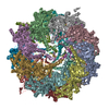
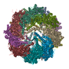
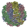
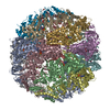
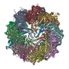
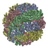
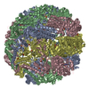
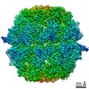
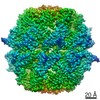
| Title | Crystal structure of the chaperonin from Thermococcus strain KS-1 (nucleotide-free form of single mutant) | ||||||
 Components Components | Thermosome alpha subunit | ||||||
 Keywords Keywords | CHAPERONE / chaperonin / thermosome | ||||||
| Function / homology |  Function and homology information Function and homology informationchaperonin ATPase / ATP-dependent protein folding chaperone / : / protein folding / ATP hydrolysis activity / ATP binding Similarity search - Function | ||||||
| Biological species |   Thermococcus sp. (archaea) Thermococcus sp. (archaea) | ||||||
| Method |  X-RAY DIFFRACTION / SYNCHROTRON / MOLECULAR REPLACEMENT / Resolution: 2.9 Å X-RAY DIFFRACTION / SYNCHROTRON / MOLECULAR REPLACEMENT / Resolution: 2.9 Å | ||||||
 Authors Authors | Shomura, Y. / Yoshida, T. / Iizuka, R. / Maruyama, T. / Yohda, M. / Miki, K. | ||||||
 Citation Citation | Journal: J.Mol.Biol. / Year: 2004 Title: Crystal Structures of the Group II Chaperonin from Thermococcus strain KS-1: Steric Hindrance by the Substituted Amino Acid, and Inter-subunit Rearrangement between Two Crystal Forms. Authors: Shomura, Y. / Yoshida, T. / Iizuka, R. / Maruyama, T. / Yohda, M. / Miki, K. #1: Journal: Acta Crystallogr.,Sect.D / Year: 2002Title: Crystallization and preliminary X-ray characterization of archaeal group II chaperonin alpha-subunit from Thermococcus strain KS-1 Authors: Shomura, Y. / Yoshida, T. / Maruyama, T. / Yohda, M. / Miki, K. | ||||||
| History |
|
- Structure visualization
| Structure viewer | Molecule: MolmilJmol/JSmol |
|---|
- Downloads & links
-Download
| PDBx/mmCIF format | 1q3r.cif.gz | 383.9 KB | Display | PDBx/mmCIF format |
|---|---|---|---|---|
| PDB format | pdb1q3r.ent.gz | 316.5 KB | Display | PDB format |
| PDBx/mmJSON format | 1q3r.json.gz | Tree view | PDBx/mmJSON format | |
| Others |  Other downloads Other downloads |
-Validation report
| Arichive directory | https://data.pdbj.org/pub/pdb/validation_reports/q3/1q3rftp://data.pdbj.org/pub/pdb/validation_reports/q3/1q3r | HTTPS FTP |
|---|
-Related structure data
| Related structure data |  1q2vSC 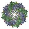 1q3qC 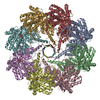 1q3sC S: Starting model for refinement C: citing same article ( |
|---|---|
| Similar structure data |

-Links
 PDBj
PDBj
- Assembly
| Deposited unit | 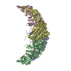
| ||||||||
|---|---|---|---|---|---|---|---|---|---|
| 1 | 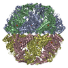
| ||||||||
| 2 | 
| ||||||||
| 3 | 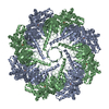
| ||||||||
| Unit cell |
| ||||||||
| Details | The biological assembly is a hexadecamer generated from the tetramer in the asymmetric unit by the operations: (-x+1, -y, z), (y+1/2, -x+1/2, z), and (-y+1/2, x-1/2, z). |
-Components
| #1: Protein | Mass: 59238.445 Da / Num. of mol.: 4 / Mutation: I125T Source method: isolated from a genetically manipulated source Source: (gene. exp.) Thermococcus sp. (archaea) / Strain: KS-1 / Gene: THSA OR CPKA / Plasmid details: pK1E-alpha1-2 / Plasmid: pET9a derivative / Species (production host): Escherichia coli / Production host:  References: UniProt: O24729, UniProt: P61112*PLUS, EC: 3.6.4.9 #2: Chemical | ChemComp-SO4 /   Mass: 96.063 Da / Num. of mol.: 4 / Source method: obtained synthetically / Formula: SO4 Mass: 96.063 Da / Num. of mol.: 4 / Source method: obtained synthetically / Formula: SO4 |
|---|
-Experimental details
-Experiment
| Experiment | Method: X-RAY DIFFRACTION / Number of used crystals: 1 |
|---|
- Sample preparation
| Crystal | Density Matthews: 3.66 Å3/Da / Density % sol: 66.37 % | |||||||||||||||||||||||||||||||||||
|---|---|---|---|---|---|---|---|---|---|---|---|---|---|---|---|---|---|---|---|---|---|---|---|---|---|---|---|---|---|---|---|---|---|---|---|---|
| Crystal grow | Temperature: 293 K / Method: vapor diffusion, sitting drop / pH: 8 Details: ammonium sulfate, potassium chloride, Tris , pH 8, VAPOR DIFFUSION, SITTING DROP, temperature 293K | |||||||||||||||||||||||||||||||||||
| Crystal grow | *PLUS Temperature: 20 ℃ / pH: 8 / Method: vapor diffusion | |||||||||||||||||||||||||||||||||||
| Components of the solutions | *PLUS
|
-Data collection
| Diffraction | Mean temperature: 90 K |
|---|---|
| Diffraction source | Source: SYNCHROTRON / Site: SPring-8  / Beamline: BL44B2 / Wavelength: 0.9 Å / Beamline: BL44B2 / Wavelength: 0.9 Å |
| Detector | Type: MARRESEARCH / Detector: CCD |
| Radiation | Monochromator: fix-exit double crystal monochromator / Protocol: SINGLE WAVELENGTH / Monochromatic (M) / Laue (L): M / Scattering type: x-ray |
| Radiation wavelength | Wavelength: 0.9 Å / Relative weight: 1 |
| Reflection | Resolution: 2.9→100 Å / Num. all: 76135 / Num. obs: 76135 / % possible obs: 95 % / Redundancy: 5.7 % / Biso Wilson estimate: 59.4 Å2 / Rsym value: 0.095 / Net I/σ(I): 12.8 |
| Reflection shell | Highest resolution: 2.9 Å / Mean I/σ(I) obs: 3.5 / Rsym value: 0.178 / % possible all: 92 |
| Reflection | *PLUS Lowest resolution: 40 Å / Num. obs: 74163 / Num. measured all: 425665 / Rmerge(I) obs: 0.095 |
| Reflection shell | *PLUS % possible obs: 92 % / Rmerge(I) obs: 0.178 |
- Processing
| Software |
| ||||||||||||||||||||||||||||||||||||||||||||||||||||||||||||||||||||||||||||||||
|---|---|---|---|---|---|---|---|---|---|---|---|---|---|---|---|---|---|---|---|---|---|---|---|---|---|---|---|---|---|---|---|---|---|---|---|---|---|---|---|---|---|---|---|---|---|---|---|---|---|---|---|---|---|---|---|---|---|---|---|---|---|---|---|---|---|---|---|---|---|---|---|---|---|---|---|---|---|---|---|---|---|
| Refinement | Method to determine structure: MOLECULAR REPLACEMENT Starting model: 1Q2V Resolution: 2.9→93.94 Å / Rfactor Rfree error: 0.004 / Data cutoff high absF: 4573193.32 / Data cutoff high rms absF: 4573193.32 / Data cutoff low absF: 0 / Isotropic thermal model: RESTRAINED / Cross valid method: THROUGHOUT / σ(F): 0 / Stereochemistry target values: Engh & Huber
| ||||||||||||||||||||||||||||||||||||||||||||||||||||||||||||||||||||||||||||||||
| Solvent computation | Solvent model: FLAT MODEL / Bsol: 19.0717 Å2 / ksol: 0.374038 e/Å3 | ||||||||||||||||||||||||||||||||||||||||||||||||||||||||||||||||||||||||||||||||
| Displacement parameters | Biso mean: 22.4 Å2
| ||||||||||||||||||||||||||||||||||||||||||||||||||||||||||||||||||||||||||||||||
| Refine analyze |
| ||||||||||||||||||||||||||||||||||||||||||||||||||||||||||||||||||||||||||||||||
| Refinement step | Cycle: LAST / Resolution: 2.9→93.94 Å
| ||||||||||||||||||||||||||||||||||||||||||||||||||||||||||||||||||||||||||||||||
| Refine LS restraints |
| ||||||||||||||||||||||||||||||||||||||||||||||||||||||||||||||||||||||||||||||||
| LS refinement shell | Resolution: 2.9→3.08 Å / Rfactor Rfree error: 0.014 / Total num. of bins used: 6
| ||||||||||||||||||||||||||||||||||||||||||||||||||||||||||||||||||||||||||||||||
| Xplor file |
| ||||||||||||||||||||||||||||||||||||||||||||||||||||||||||||||||||||||||||||||||
| Refinement | *PLUS Highest resolution: 2.9 Å / Lowest resolution: 93.9 Å | ||||||||||||||||||||||||||||||||||||||||||||||||||||||||||||||||||||||||||||||||
| Solvent computation | *PLUS | ||||||||||||||||||||||||||||||||||||||||||||||||||||||||||||||||||||||||||||||||
| Displacement parameters | *PLUS | ||||||||||||||||||||||||||||||||||||||||||||||||||||||||||||||||||||||||||||||||
| Refine LS restraints | *PLUS
|
