- PDB-3kkv: Structure of PKA with a protein Kinase B-selective inhibitor. -
+
Open data
ID or keywords:
Loading...
-
Basic information
Entry
Database: PDB / ID: 3kkv
Title
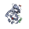
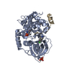
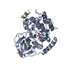
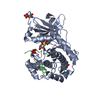
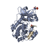
Structure of PKA with a protein Kinase B-selective inhibitor.
Components
PKI kinase inhibitor
cAMP-dependent protein kinase catalytic subunit alpha
Keywords
TRANSFERASE/TRANSFERASE INHIBITOR / protein kinase / TRANSFERASE-TRANSFERASE INHIBITOR complex
Function / homology
Function and homology information
CD209 (DC-SIGN) signaling / HDL assembly / Regulation of insulin secretion / Rap1 signalling / Ion homeostasis / PKA activation in glucagon signalling / DARPP-32 events / CREB1 phosphorylation through the activation of Adenylate Cyclase / GPER1 signaling / Factors involved in megakaryocyte development and platelet production ...CD209 (DC-SIGN) signaling / HDL assembly / Regulation of insulin secretion / Rap1 signalling / Ion homeostasis / PKA activation in glucagon signalling / DARPP-32 events / CREB1 phosphorylation through the activation of Adenylate Cyclase / GPER1 signaling / Factors involved in megakaryocyte development and platelet production / Loss of Nlp from mitotic centrosomes / Recruitment of mitotic centrosome proteins and complexes / Loss of proteins required for interphase microtubule organization from the centrosome / Anchoring of the basal body to the plasma membrane / RET signaling / AURKA Activation by TPX2 / Interleukin-3, Interleukin-5 and GM-CSF signaling / Recruitment of NuMA to mitotic centrosomes / VEGFA-VEGFR2 Pathway / PKA activation / MAPK6/MAPK4 signaling / GLI3 is processed to GLI3R by the proteasome / Regulation of PLK1 Activity at G2/M Transition / Hedgehog 'off' state / histone H1-4S35 kinase activity / cAMP-dependent protein kinase / regulation of protein processing / cAMP-dependent protein kinase activity / cellular response to parathyroid hormone stimulus / protein localization to lipid droplet / regulation of bicellular tight junction assembly / cAMP-dependent protein kinase complex / dorsal/ventral neural tube patterning / interleukin-2-mediated signaling pathway / Mitochondrial protein degradation / regulation of osteoblast differentiation / Glucagon-like Peptide-1 (GLP1) regulates insulin secretion / sperm capacitation / cellular response to cold / High laminar flow shear stress activates signaling by PIEZO1 and PECAM1:CDH5:KDR in endothelial cells / Vasopressin regulates renal water homeostasis via Aquaporins / protein kinase A regulatory subunit binding / negative regulation of glycolytic process through fructose-6-phosphate / ciliary base / intracellular potassium ion homeostasis / mesoderm formation / TORC1 signaling / plasma membrane raft / axoneme / sperm flagellum / postsynaptic modulation of chemical synaptic transmission / negative regulation of TORC1 signaling / protein serine/threonine/tyrosine kinase activity / regulation of proteasomal protein catabolic process / protein export from nucleus / cellular response to glucagon stimulus / cellular response to nutrient levels / positive regulation of phagocytosis / positive regulation of gluconeogenesis / acrosomal vesicle / positive regulation of protein export from nucleus / neuromuscular junction / negative regulation of smoothened signaling pathway / neural tube closure / cellular response to glucose stimulus / positive regulation of cholesterol biosynthetic process / mRNA processing / adenylate cyclase-inhibiting G protein-coupled receptor signaling pathway / positive regulation of insulin secretion / manganese ion binding / cellular response to heat / adenylate cyclase-activating G protein-coupled receptor signaling pathway / protein kinase activity / regulation of cell cycle / nuclear speck / postsynapse / protein domain specific binding / protein serine kinase activity / protein serine/threonine kinase activity / ubiquitin protein ligase binding / centrosome / protein kinase binding / perinuclear region of cytoplasm / glutamatergic synapse / magnesium ion binding / mitochondrion / ATP binding / nucleus / cytoplasm / cytosol Similarity search - Function
cAMP-dependent protein kinase catalytic subunit / Extension to Ser/Thr-type protein kinases / AGC-kinase, C-terminal / AGC-kinase C-terminal domain profile. / Phosphorylase Kinase; domain 1 / Phosphorylase Kinase; domain 1 / Transferase(Phosphotransferase) domain 1 / Transferase(Phosphotransferase); domain 1 / Serine/threonine-protein kinase, active site / Serine/Threonine protein kinases active-site signature. ...cAMP-dependent protein kinase catalytic subunit / Extension to Ser/Thr-type protein kinases / AGC-kinase, C-terminal / AGC-kinase C-terminal domain profile. / Phosphorylase Kinase; domain 1 / Phosphorylase Kinase; domain 1 / Transferase(Phosphotransferase) domain 1 / Transferase(Phosphotransferase); domain 1 / Serine/threonine-protein kinase, active site / Serine/Threonine protein kinases active-site signature. / Protein kinase domain / Serine/Threonine protein kinases, catalytic domain / Protein kinase, ATP binding site / Protein kinases ATP-binding region signature. / Protein kinase domain profile. / Protein kinase domain / Protein kinase-like domain superfamily / 2-Layer Sandwich / Orthogonal Bundle / Mainly Alpha / Alpha Beta Similarity search - Domain/homology
Mass: 18.015 Da / Num. of mol.: 251 / Source method: isolated from a natural source / Formula: H2O
Has protein modification
Y
-
Experimental details
-
Experiment
Experiment
Method: X-RAY DIFFRACTION / Number of used crystals: 1
-
Sample preparation
Crystal
Density Matthews: 2.57 Å3/Da / Density % sol: 52.16 %
Crystal grow
Temperature: 294 K / Method: vapor diffusion, sitting drop / pH: 6.4 Details: Protein stock was at 20mg/ml in 25mM Mes-Bis Tris, pH 6.4, 75mM LiCl, 0.1M EDTA, 1mM DTT. The final concentration of inhibitor in the protein solution was 1mM from a 100mM stock solution of ...Details: Protein stock was at 20mg/ml in 25mM Mes-Bis Tris, pH 6.4, 75mM LiCl, 0.1M EDTA, 1mM DTT. The final concentration of inhibitor in the protein solution was 1mM from a 100mM stock solution of compound dissolved in DMSO.The protein was reacted with inhibitor on ice for 6 hours prior to crystallization.The final concentration of PKI in the protein solution was 2mM from stock 100mM dissolved in water. The final concentration of MEGA8 in the protein solution was 1.5mM from 225mM stock diluted from 790mM in the Hampton additive kit with water. Two microliters of protein were added to 0.35 microliters of 10% glycerol in buffer and put in sitting drops over 15% methanol diluted with water at 4C. Cryo was 100mM MES pH6.4, 100mM LiCl, 25% MeOH, 20% MPD., VAPOR DIFFUSION, SITTING DROP, temperature 294K
Monochromator: downward deflecting liquid nitrogen cooled Si(111) double-crystal system monochromator designed and developed by Daresbury Laboratory (UK). Protocol: SINGLE WAVELENGTH / Monochromatic (M) / Laue (L): M / Scattering type: x-ray
Radiation wavelength
Wavelength: 1 Å / Relative weight: 1
Reflection
Resolution: 1.8→50 Å / Num. all: 41841 / Num. obs: 41799 / % possible obs: 99.9 % / Observed criterion σ(F): 2 / Observed criterion σ(I): 2 / Biso Wilson estimate: 24 Å2 / Net I/σ(I): 37
Reflection shell
Resolution: 1.8→1.86 Å / % possible all: 99.6
-
Processing
Software
Name
Version
Classification
ADSC
Quantum
datacollection
AMoRE
phasing
PHENIX
(phenix.refine: 1.5_2)
refinement
HKL-2000
datareduction
HKL-2000
datascaling
Refinement
Method to determine structure: MOLECULAR REPLACEMENT / Resolution: 1.8→36.206 Å / SU ML: 0.22 / σ(F): 1.9 / Stereochemistry target values: ML
Rfactor
Num. reflection
% reflection
Selection details
Rfree
0.2252
2108
5.05 %
Random
Rwork
0.2017
-
-
-
all
0.2028
41840
-
-
obs
0.2028
41735
99.88 %
-
Solvent computation
Shrinkage radii: 0.9 Å / VDW probe radii: 1.11 Å / Solvent model: FLAT BULK SOLVENT MODEL / Bsol: 40.067 Å2 / ksol: 0.328 e/Å3
Refinement step
Cycle: LAST / Resolution: 1.8→36.206 Å
Protein
Nucleic acid
Ligand
Solvent
Total
Num. atoms
2956
0
35
251
3242
Refine LS restraints
Refine-ID
Type
Dev ideal
Number
X-RAY DIFFRACTION
f_bond_d
0.001
3071
X-RAY DIFFRACTION
f_angle_d
0.59
4150
X-RAY DIFFRACTION
f_dihedral_angle_d
13.079
1122
X-RAY DIFFRACTION
f_chiral_restr
0.034
429
X-RAY DIFFRACTION
f_plane_restr
0.002
540
LS refinement shell
Resolution (Å)
Rfactor Rfree
Num. reflection Rfree
Rfactor Rwork
Num. reflection Rwork
Refine-ID
% reflection obs (%)
1.8-1.8421
0.2573
155
0.2332
2567
X-RAY DIFFRACTION
99
1.8421-1.8881
0.2603
134
0.2223
2589
X-RAY DIFFRACTION
100
1.8881-1.9392
0.2211
132
0.2149
2635
X-RAY DIFFRACTION
100
1.9392-1.9962
0.2447
137
0.21
2599
X-RAY DIFFRACTION
100
1.9962-2.0607
0.2097
135
0.2033
2621
X-RAY DIFFRACTION
100
2.0607-2.1343
0.2365
154
0.1945
2601
X-RAY DIFFRACTION
100
2.1343-2.2197
0.2631
136
0.2022
2613
X-RAY DIFFRACTION
100
2.2197-2.3208
0.2623
134
0.2028
2637
X-RAY DIFFRACTION
100
2.3208-2.4431
0.2189
141
0.207
2637
X-RAY DIFFRACTION
100
2.4431-2.5961
0.2605
139
0.2047
2637
X-RAY DIFFRACTION
100
2.5961-2.7965
0.2542
139
0.2288
2634
X-RAY DIFFRACTION
100
2.7965-3.0778
0.235
137
0.2202
2667
X-RAY DIFFRACTION
100
3.0778-3.5228
0.2124
140
0.2074
2670
X-RAY DIFFRACTION
100
3.5228-4.4371
0.2045
160
0.1704
2698
X-RAY DIFFRACTION
100
4.4371-36.2128
0.1789
135
0.1718
2822
X-RAY DIFFRACTION
99
Refinement TLS params.
Refine-ID: X-RAY DIFFRACTION
ID
Method
L11 (°2)
L12 (°2)
L13 (°2)
L22 (°2)
L23 (°2)
L33 (°2)
S11 (Å °)
S12 (Å °)
S13 (Å °)
S21 (Å °)
S22 (Å °)
S23 (Å °)
S31 (Å °)
S32 (Å °)
S33 (Å °)
T11 (Å2)
T12 (Å2)
T13 (Å2)
T22 (Å2)
T23 (Å2)
T33 (Å2)
Origin x (Å)
Origin y (Å)
Origin z (Å)
1
refined
0.8299
0.1049
0.0037
0.9053
-0.41
1.4669
0.0024
0.0573
-0.0042
0.037
0.0278
0.0781
-0.1238
-0.1396
-0.0247
0.1095
0.0235
0.0038
0.1558
-0.0188
0.1357
-13.6306
0.9107
-8.5711
2
0.3703
-0.107
0.0965
0.3907
-0.4832
0.8505
-0.1005
0.0782
-0.0489
0.1829
-0.0804
-0.0902
-0.0625
0.162
0.106
0.2452
0.0197
0.0015
0.2167
-0.002
0.1795
Refinement TLS group
ID
Refine-ID
Refine TLS-ID
Selection details
Auth asym-ID
Auth seq-ID
1
X-RAY DIFFRACTION
1
chainAandresid8:350
A
8 - 350
2
X-RAY DIFFRACTION
2
chainIandresid5:24
I
5 - 24
+
About Yorodumi
-
News
-
Feb 9, 2022. New format data for meta-information of EMDB entries
New format data for meta-information of EMDB entries
Version 3 of the EMDB header file is now the official format.
The previous official version 1.9 will be removed from the archive.
In the structure databanks used in Yorodumi, some data are registered as the other names, "COVID-19 virus" and "2019-nCoV". Here are the details of the virus and the list of structure data.
Jan 31, 2019. EMDB accession codes are about to change! (news from PDBe EMDB page)
EMDB accession codes are about to change! (news from PDBe EMDB page)
The allocation of 4 digits for EMDB accession codes will soon come to an end. Whilst these codes will remain in use, new EMDB accession codes will include an additional digit and will expand incrementally as the available range of codes is exhausted. The current 4-digit format prefixed with “EMD-” (i.e. EMD-XXXX) will advance to a 5-digit format (i.e. EMD-XXXXX), and so on. It is currently estimated that the 4-digit codes will be depleted around Spring 2019, at which point the 5-digit format will come into force.
The EM Navigator/Yorodumi systems omit the EMD- prefix.
Related info.:Q: What is EMD? / ID/Accession-code notation in Yorodumi/EM Navigator
Yorodumi is a browser for structure data from EMDB, PDB, SASBDB, etc.
This page is also the successor to EM Navigator detail page, and also detail information page/front-end page for Omokage search.
The word "yorodu" (or yorozu) is an old Japanese word meaning "ten thousand". "mi" (miru) is to see.
Related info.:EMDB / PDB / SASBDB / Comparison of 3 databanks / Yorodumi Search / Aug 31, 2016. New EM Navigator & Yorodumi / Yorodumi Papers / Jmol/JSmol / Function and homology information / Changes in new EM Navigator and Yorodumi
 Movie
Movie Controller


 Open data
 Basic information
 Components
 Keywords
 Function and homology information
 X-RAY DIFFRACTION /
  Authors
 Citation
 Structure visualization
 Downloads & links
 Other downloads


 PDBj
PDBj


 Assembly
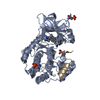


 Mass: 463.530 Da / Num. of mol.: 1 / Source method: obtained synthetically / Formula: C28H25N5O2
 Mass: 18.015 Da / Num. of mol.: 251 / Source method: isolated from a natural source / Formula: H2O
 Sample preparation
 / Beamline: 17-ID / Wavelength: 1 Å
 Processing