 Movie
Movie Controller

[English] 日本語
 Yorodumi
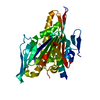
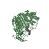
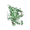
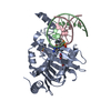
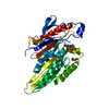
- PDB-3kar: THE MOTOR DOMAIN OF KINESIN-LIKE PROTEIN KAR3, A SACCHAROMYCES CE... -
+ Open data

- Basic information
| Entry | Database: PDB / ID: 3kar | ||||||
|---|---|---|---|---|---|---|---|
| Title | THE MOTOR DOMAIN OF KINESIN-LIKE PROTEIN KAR3, A SACCHAROMYCES CEREVISIAE KINESIN-RELATED PROTEIN | ||||||
 Components Components | KINESIN-LIKE PROTEIN KAR3 | ||||||
 Keywords Keywords | CONTRACTILE PROTEIN / KAR3 / KINESIN-RELATED PROTEIN / MOTOR PROTEIN / ATPASE / P-LOOP / MICROTUBULE BINDING PROTEIN | ||||||
| Function / homology |  Function and homology information Function and homology informationminus-end-directed kinesin ATPase / nuclear migration involved in conjugation with cellular fusion / protein transport along microtubule to mitotic spindle pole body / karyogamy involved in conjugation with cellular fusion / mitotic spindle pole body / mitotic spindle midzone assembly / spindle pole body / minus-end-directed microtubule motor activity / mitotic sister chromatid cohesion / microtubule-based process ...minus-end-directed kinesin ATPase / nuclear migration involved in conjugation with cellular fusion / protein transport along microtubule to mitotic spindle pole body / karyogamy involved in conjugation with cellular fusion / mitotic spindle pole body / mitotic spindle midzone assembly / spindle pole body / minus-end-directed microtubule motor activity / mitotic sister chromatid cohesion / microtubule-based process / cytoplasmic microtubule / ERAD pathway / regulation of mitotic spindle organization / meiotic cell cycle / spindle pole / mitotic cell cycle / chromosome / microtubule cytoskeleton / microtubule binding / microtubule / cell division / ATP hydrolysis activity / ATP binding / nucleus Similarity search - Function | ||||||
| Biological species |  | ||||||
| Method |  X-RAY DIFFRACTION / MIR / Resolution: 2.3 Å X-RAY DIFFRACTION / MIR / Resolution: 2.3 Å | ||||||
 Authors Authors | Gulick, A.M. / Song, H. / Endow, S. / Rayment, I. | ||||||
 Citation Citation | Journal: Biochemistry / Year: 1998 Title: X-ray crystal structure of the yeast Kar3 motor domain complexed with Mg.ADP to 2.3 A resolution. Authors: Gulick, A.M. / Song, H. / Endow, S.A. / Rayment, I. | ||||||
| History |
|
- Structure visualization
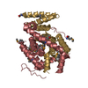
| Structure viewer | Molecule: MolmilJmol/JSmol |
|---|
- Downloads & links
-Download
| PDBx/mmCIF format | 3kar.cif.gz | 81 KB | Display | PDBx/mmCIF format |
|---|---|---|---|---|
| PDB format | pdb3kar.ent.gz | 57.9 KB | Display | PDB format |
| PDBx/mmJSON format | 3kar.json.gz | Tree view | PDBx/mmJSON format | |
| Others |  Other downloads Other downloads |
-Validation report
| Arichive directory | https://data.pdbj.org/pub/pdb/validation_reports/ka/3karftp://data.pdbj.org/pub/pdb/validation_reports/ka/3kar | HTTPS FTP |
|---|
-Related structure data
| Similar structure data |
|---|


-Links
 PDBj
PDBj


- Assembly
| Deposited unit | 
| ||||||||
|---|---|---|---|---|---|---|---|---|---|
| 1 |
| ||||||||
| Unit cell |
|
-Components
| #1: Protein | Mass: 38806.539 Da / Num. of mol.: 1 / Fragment: MOTOR DOMAIN, RESIDUES 383 - 722 / Mutation: L383M Source method: isolated from a genetically manipulated source Details: GENETICALLY TRUNCATED MOTOR DOMAIN OF SACCHAROMYCES CEREVISIAE MOTOR PROTEIN KAR3 LIGANDS MG2 AND ADP Source: (gene. exp.) Gene: KAR3 / Plasmid: PMW-KAR3 / Production host:  |
|---|---|
| #2: Chemical | ChemComp-MG /   Mass: 24.305 Da / Num. of mol.: 1 / Source method: obtained synthetically / Formula: Mg Mass: 24.305 Da / Num. of mol.: 1 / Source method: obtained synthetically / Formula: Mg |
| #3: Chemical | ChemComp-ADP /   Mass: 427.201 Da / Num. of mol.: 1 / Source method: obtained synthetically / Formula: C10H15N5O10P2 / Comment: ADP, energy-carrying molecule*YM Mass: 427.201 Da / Num. of mol.: 1 / Source method: obtained synthetically / Formula: C10H15N5O10P2 / Comment: ADP, energy-carrying molecule*YM |
| #4: Water | ChemComp-HOH /  Mass: 18.015 Da / Num. of mol.: 105 / Source method: isolated from a natural source / Formula: H2O Mass: 18.015 Da / Num. of mol.: 105 / Source method: isolated from a natural source / Formula: H2O |
-Experimental details
-Experiment
| Experiment | Method: X-RAY DIFFRACTION / Number of used crystals: 1 |
|---|
- Sample preparation
| Crystal | Density Matthews: 2.14 Å3/Da / Density % sol: 42 % | ||||||||||||||||||||||||||||||||||||
|---|---|---|---|---|---|---|---|---|---|---|---|---|---|---|---|---|---|---|---|---|---|---|---|---|---|---|---|---|---|---|---|---|---|---|---|---|---|
| Crystal grow | pH: 7 Details: 11 % MEPEG 2000, 50 MM NACL, 1% ETHYLENE GLYCOL, 1 MM MG ADP, pH 7. | ||||||||||||||||||||||||||||||||||||
| Crystal grow | *PLUS pH: 7 / Method: batch method | ||||||||||||||||||||||||||||||||||||
| Components of the solutions | *PLUS
|
-Data collection
| Diffraction | Mean temperature: 269 K |
|---|---|
| Diffraction source | Source: ROTATING ANODE / Type: RIGAKU RUH2R / Wavelength: 1.5418 |
| Detector | Type: SIEMENS HI-STAR / Detector: AREA DETECTOR / Date: Apr 9, 1997 |
| Radiation | Monochromatic (M) / Laue (L): M / Scattering type: x-ray |
| Radiation wavelength | Wavelength: 1.5418 Å / Relative weight: 1 |
| Reflection | Resolution: 2.3→30 Å / Num. obs: 22220 / % possible obs: 93 % / Redundancy: 2 % / Rmerge(I) obs: 0.034 |
| Reflection shell | Resolution: 2.3→2.4 Å / Rmerge(I) obs: 0.074 / % possible all: 85 |
| Reflection | *PLUS Num. measured all: 43898 |
| Reflection shell | *PLUS % possible obs: 84.7 % |
- Processing
| Software |
| ||||||||||||||||||||||||||||||
|---|---|---|---|---|---|---|---|---|---|---|---|---|---|---|---|---|---|---|---|---|---|---|---|---|---|---|---|---|---|---|---|
| Refinement | Method to determine structure: MIR / Resolution: 2.3→30 Å
| ||||||||||||||||||||||||||||||
| Refinement step | Cycle: LAST / Resolution: 2.3→30 Å
| ||||||||||||||||||||||||||||||
| Refine LS restraints |
| ||||||||||||||||||||||||||||||
| Software | *PLUS Name: TNT / Classification: refinement | ||||||||||||||||||||||||||||||
| Refinement | *PLUS Rfactor all: 0.175 | ||||||||||||||||||||||||||||||
| Solvent computation | *PLUS | ||||||||||||||||||||||||||||||
| Displacement parameters | *PLUS | ||||||||||||||||||||||||||||||
| Refine LS restraints | *PLUS
|
