| Entry | Database: PDB / ID: 3zns
|
|---|
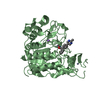
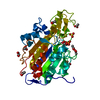
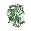
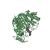
| Title | HDAC7 bound with TFMO inhibitor tmp942 |
|---|
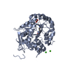
 Components Components | HISTONE DEACETYLASE 7 |
|---|
 Keywords Keywords | HYDROLASE / ZBG / MBG |
|---|
| Function / homology |  Function and homology information Function and homology information
regulation of mRNA processing / protein deacetylation / histone deacetylase activity, hydrolytic mechanism / histone deacetylase / cell-cell junction assembly / SUMO transferase activity / negative regulation of non-canonical NF-kappaB signal transduction / histone deacetylase activity / protein lysine deacetylase activity / Hydrolases; Acting on carbon-nitrogen bonds, other than peptide bonds; In linear amides ...regulation of mRNA processing / protein deacetylation / histone deacetylase activity, hydrolytic mechanism / histone deacetylase / cell-cell junction assembly / SUMO transferase activity / negative regulation of non-canonical NF-kappaB signal transduction / histone deacetylase activity / protein lysine deacetylase activity / Hydrolases; Acting on carbon-nitrogen bonds, other than peptide bonds; In linear amides / positive regulation of cell migration involved in sprouting angiogenesis / negative regulation of interleukin-2 production / Notch-HLH transcription pathway / histone deacetylase complex / protein sumoylation / vasculogenesis / negative regulation of osteoblast differentiation / SUMOylation of DNA damage response and repair proteins / 14-3-3 protein binding / epigenetic regulation of gene expression / protein kinase C binding / Regulation of PTEN gene transcription / NOTCH1 Intracellular Domain Regulates Transcription / Constitutive Signaling by NOTCH1 PEST Domain Mutants / Constitutive Signaling by NOTCH1 HD+PEST Domain Mutants / transcription corepressor activity / DNA-binding transcription factor binding / chromatin binding / protein kinase binding / negative regulation of transcription by RNA polymerase II / nucleoplasm / metal ion binding / nucleus / cytoplasm / cytosolSimilarity search - Function : / Histone deacetylase domain / Arginase; Chain A / Histone deacetylase family / Histone deacetylase domain / Histone deacetylase domain superfamily / Histone deacetylase domain / Ureohydrolase domain superfamily / 3-Layer(aba) Sandwich / Alpha BetaSimilarity search - Domain/homology |
|---|
| Biological species |  HOMO SAPIENS (human) HOMO SAPIENS (human) |
|---|
| Method |  X-RAY DIFFRACTION / SYNCHROTRON / MOLECULAR REPLACEMENT / Resolution: 2.45 Å X-RAY DIFFRACTION / SYNCHROTRON / MOLECULAR REPLACEMENT / Resolution: 2.45 Å |
|---|
 Authors Authors | Lobera, M. / Madauss, K.P. / Pohlhaus, D.T. / Trump, R.P. / Nolan, M.A. |
|---|
 Citation Citation | Journal: Nat.Chem.Biol. / Year: 2013
Title: Selective Class Iia Histone Deacetylase Inhibition Via a Non-Chelating Zinc Binding Group
Authors: Lobera, M. / Madauss, K.P. / Pohlhaus, D.T. / Wright, Q.G. / Trocha, M. / Schmidt, D.R. / Baloglu, E. / Trump, R.P. / Head, M.S. / Hofmann, G.A. / Murray-Thompson, M. / Schwartz, B. / ...Authors: Lobera, M. / Madauss, K.P. / Pohlhaus, D.T. / Wright, Q.G. / Trocha, M. / Schmidt, D.R. / Baloglu, E. / Trump, R.P. / Head, M.S. / Hofmann, G.A. / Murray-Thompson, M. / Schwartz, B. / Chakravorty, S. / Wu, Z. / Mander, P.K. / Kruidenier, L. / Reid, R.A. / Burkhart, W. / Turunen, B.J. / Rong, J.X. / Wagner, C. / Moyer, M.B. / Wells, C. / Hong, X. / Moore, J.T. / Williams, J.D. / Soler, D. / Ghosh, S. / Nolan, M.A. |
|---|
| History | | Deposition | Feb 15, 2013 | Deposition site: PDBE / Processing site: PDBE |
|---|
| Revision 1.0 | Mar 27, 2013 | Provider: repository / Type: Initial release |
|---|
| Revision 1.1 | Apr 3, 2013 | Group: Database references |
|---|
| Revision 1.2 | May 1, 2013 | Group: Database references |
|---|
| Revision 1.3 | May 8, 2024 | Group: Data collection / Database references ...Data collection / Database references / Derived calculations / Other
Category: chem_comp_atom / chem_comp_bond ...chem_comp_atom / chem_comp_bond / database_2 / pdbx_database_status / pdbx_struct_conn_angle / struct_conn / struct_site
Item: _database_2.pdbx_DOI / _database_2.pdbx_database_accession ..._database_2.pdbx_DOI / _database_2.pdbx_database_accession / _pdbx_database_status.status_code_sf / _pdbx_struct_conn_angle.ptnr1_auth_comp_id / _pdbx_struct_conn_angle.ptnr1_auth_seq_id / _pdbx_struct_conn_angle.ptnr1_label_asym_id / _pdbx_struct_conn_angle.ptnr1_label_atom_id / _pdbx_struct_conn_angle.ptnr1_label_comp_id / _pdbx_struct_conn_angle.ptnr1_label_seq_id / _pdbx_struct_conn_angle.ptnr3_auth_comp_id / _pdbx_struct_conn_angle.ptnr3_auth_seq_id / _pdbx_struct_conn_angle.ptnr3_label_asym_id / _pdbx_struct_conn_angle.ptnr3_label_atom_id / _pdbx_struct_conn_angle.ptnr3_label_comp_id / _pdbx_struct_conn_angle.ptnr3_label_seq_id / _pdbx_struct_conn_angle.value / _struct_conn.pdbx_dist_value / _struct_conn.ptnr1_auth_comp_id / _struct_conn.ptnr1_auth_seq_id / _struct_conn.ptnr1_label_asym_id / _struct_conn.ptnr1_label_atom_id / _struct_conn.ptnr1_label_comp_id / _struct_conn.ptnr1_label_seq_id / _struct_conn.ptnr2_auth_comp_id / _struct_conn.ptnr2_auth_seq_id / _struct_conn.ptnr2_label_asym_id / _struct_conn.ptnr2_label_atom_id / _struct_conn.ptnr2_label_comp_id / _struct_conn.ptnr2_label_seq_id / _struct_site.pdbx_auth_asym_id / _struct_site.pdbx_auth_comp_id / _struct_site.pdbx_auth_seq_id |
|---|
|
|---|
 Movie
Movie Controller


 Open data
 Basic information
 Structure visualization
 Downloads & links
 Other downloads
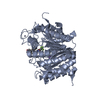


 PDBj
PDBj


 Assembly
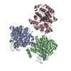


 Mass: 65.409 Da / Num. of mol.: 6 / Source method: obtained synthetically / Formula: Zn
 Mass: 39.098 Da / Num. of mol.: 6 / Source method: obtained synthetically / Formula: K
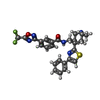
 Mass: 527.561 Da / Num. of mol.: 3 / Source method: obtained synthetically / Formula: C26H24F3N5O2S
 Mass: 18.015 Da / Num. of mol.: 133 / Source method: isolated from a natural source / Formula: H2O
 Sample preparation
 / Beamline: 21-ID-G / Wavelength: 0.97
  Processing