 Movie
Movie Controller

[English] 日本語
 Yorodumi
- PDB-3k75: X-ray crystal structure of reduced XRCC1 bound to DNA pol beta ca... -
+ Open data

- Basic information
| Entry | Database: PDB / ID: 3k75 | ||||||
|---|---|---|---|---|---|---|---|
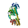
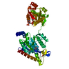
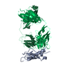
| Title | X-ray crystal structure of reduced XRCC1 bound to DNA pol beta catalytic domain | ||||||
 Components Components |
| ||||||
 Keywords Keywords | DNA BINDING PROTEIN / allosteric disulfide / XRCC1 / pol beta / DNA damage / DNA repair / Nucleus / Phosphoprotein / DNA replication / DNA synthesis / DNA-binding / DNA-directed DNA polymerase / Lyase / Magnesium / Metal-binding / Methylation / Nucleotidyltransferase / Transferase / DNA-BINDING PROTEIN | ||||||
| Function / homology |  Function and homology information Function and homology informationoxidized DNA binding / Resolution of AP sites via the multiple-nucleotide patch replacement pathway / Resolution of AP sites via the single-nucleotide replacement pathway / APEX1-Independent Resolution of AP Sites via the Single Nucleotide Replacement Pathway / PCNA-Dependent Long Patch Base Excision Repair / Abasic sugar-phosphate removal via the single-nucleotide replacement pathway / POLB-Dependent Long Patch Base Excision Repair / telomeric DNA-containing double minutes formation / ERCC4-ERCC1 complex / negative regulation of protection from non-homologous end joining at telomere ...oxidized DNA binding / Resolution of AP sites via the multiple-nucleotide patch replacement pathway / Resolution of AP sites via the single-nucleotide replacement pathway / APEX1-Independent Resolution of AP Sites via the Single Nucleotide Replacement Pathway / PCNA-Dependent Long Patch Base Excision Repair / Abasic sugar-phosphate removal via the single-nucleotide replacement pathway / POLB-Dependent Long Patch Base Excision Repair / telomeric DNA-containing double minutes formation / ERCC4-ERCC1 complex / negative regulation of protection from non-homologous end joining at telomere / ADP-D-ribose modification-dependent protein binding / negative regulation of protein ADP-ribosylation / somatic diversification of immunoglobulins / Ub-specific processing proteases / poly-ADP-D-ribose binding / regulation of base-excision repair / single strand break repair / HDR through MMEJ (alt-NHEJ) / 3' overhang single-stranded DNA endonuclease activity / response to hydroperoxide / Resolution of AP sites via the single-nucleotide replacement pathway / immunoglobulin heavy chain V-D-J recombination / APEX1-Independent Resolution of AP Sites via the Single Nucleotide Replacement Pathway / Lyases; Carbon-oxygen lyases; Other carbon-oxygen lyases / pyrimidine dimer repair / homeostasis of number of cells / 5'-deoxyribose-5-phosphate lyase activity / response to hyperoxia / lymph node development / salivary gland morphogenesis / somatic hypermutation of immunoglobulin genes / spleen development / site of DNA damage / base-excision repair, gap-filling / class I DNA-(apurinic or apyrimidinic site) endonuclease activity / DNA-(apurinic or apyrimidinic site) lyase / response to gamma radiation / Gap-filling DNA repair synthesis and ligation in GG-NER / spindle microtubule / hippocampus development / base-excision repair / double-strand break repair via nonhomologous end joining / intrinsic apoptotic signaling pathway in response to DNA damage / Gap-filling DNA repair synthesis and ligation in TC-NER / double-strand break repair / neuron apoptotic process / DNA-directed DNA polymerase / microtubule binding / in utero embryonic development / damaged DNA binding / microtubule / response to ethanol / DNA-directed DNA polymerase activity / chromosome, telomeric region / DNA replication / lyase activity / inflammatory response / apoptotic process / DNA damage response / chromatin / nucleolus / enzyme binding / protein-containing complex / DNA binding / nucleoplasm / metal ion binding / nucleus / cytoplasm Similarity search - Function | ||||||
| Biological species |  Homo sapiens (human) Homo sapiens (human) | ||||||
| Method |  X-RAY DIFFRACTION / MIR / Resolution: 2.95 Å X-RAY DIFFRACTION / MIR / Resolution: 2.95 Å | ||||||
 Authors Authors | Cuneo, M.J. / London, R.E. | ||||||
 Citation Citation | Journal: Proc.Natl.Acad.Sci.USA / Year: 2010 Title: Oxidation state of the XRCC1 N-terminal domain regulates DNA polymerase beta binding affinity. Authors: Cuneo, M.J. / London, R.E. | ||||||
| History |
|
- Structure visualization
| Structure viewer | Molecule: MolmilJmol/JSmol |
|---|
- Downloads & links
-Download
| PDBx/mmCIF format | 3k75.cif.gz | 320.7 KB | Display | PDBx/mmCIF format |
|---|---|---|---|---|
| PDB format | pdb3k75.ent.gz | 263.5 KB | Display | PDB format |
| PDBx/mmJSON format | 3k75.json.gz | Tree view | PDBx/mmJSON format | |
| Others |  Other downloads Other downloads |
-Validation report
| Arichive directory | https://data.pdbj.org/pub/pdb/validation_reports/k7/3k75ftp://data.pdbj.org/pub/pdb/validation_reports/k7/3k75 | HTTPS FTP |
|---|
-Related structure data
| Related structure data |  3k77C  3lqcC  3k76 C: citing same article ( |
|---|---|
| Similar structure data |

-Links
 PDBj
PDBj


- Assembly
| Deposited unit | 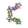
| ||||||||||||||||||||||||||||||||||||||||||||||||||||||||||||||||||||
|---|---|---|---|---|---|---|---|---|---|---|---|---|---|---|---|---|---|---|---|---|---|---|---|---|---|---|---|---|---|---|---|---|---|---|---|---|---|---|---|---|---|---|---|---|---|---|---|---|---|---|---|---|---|---|---|---|---|---|---|---|---|---|---|---|---|---|---|---|---|
| 1 | 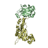
| ||||||||||||||||||||||||||||||||||||||||||||||||||||||||||||||||||||
| 2 | 
| ||||||||||||||||||||||||||||||||||||||||||||||||||||||||||||||||||||
| Unit cell |
| ||||||||||||||||||||||||||||||||||||||||||||||||||||||||||||||||||||
| Noncrystallographic symmetry (NCS) | NCS domain:
NCS domain segments: Component-ID: 1 / Refine code: 4
NCS ensembles :
|
-Components
| #1: Protein | Mass: 21060.527 Da / Num. of mol.: 2 / Fragment: N-terminal domain (UNP residues 1 to 183) Source method: isolated from a genetically manipulated source Source: (gene. exp.) Homo sapiens (human) / Strain: Human / Gene: XRCC1 / Plasmid: pET21a / Production host:  #2: Protein | Mass: 29336.111 Da / Num. of mol.: 2 / Fragment: UNP residues 91 to 335 Source method: isolated from a genetically manipulated source Source: (gene. exp.) References: UniProt: P06766, DNA-directed DNA polymerase, Lyases; Carbon-oxygen lyases; Other carbon-oxygen lyases |
|---|
-Experimental details
-Experiment
| Experiment | Method: X-RAY DIFFRACTION / Number of used crystals: 1 |
|---|
- Sample preparation
| Crystal | Density Matthews: 2.24 Å3/Da / Density % sol: 45.18 % |
|---|---|
| Crystal grow | Temperature: 298 K / Method: vapor diffusion, hanging drop / pH: 7.5 Details: 20-25% PEG 3350, 0.2-0.3M Tri-potassium citrate, pH 7.5, VAPOR DIFFUSION, HANGING DROP, temperature 298K |
-Data collection
| Diffraction | Mean temperature: 100 K |
|---|---|
| Diffraction source | Source: ROTATING ANODE / Type: RIGAKU MICROMAX-007 HF / Wavelength: 1.5418 Å |
| Detector | Type: RIGAKU SATURN 92 / Detector: CCD / Date: Sep 30, 2008 |
| Radiation | Protocol: SINGLE WAVELENGTH / Monochromatic (M) / Laue (L): M / Scattering type: x-ray |
| Radiation wavelength | Wavelength: 1.5418 Å / Relative weight: 1 |
| Reflection | Resolution: 2.95→25 Å / Num. obs: 19123 / % possible obs: 98.7 % / Observed criterion σ(F): 0 / Observed criterion σ(I): -3 / Biso Wilson estimate: 41.334 Å2 / Rmerge(I) obs: 0.061 / Net I/σ(I): 17.7 |
| Reflection shell | Resolution: 2.95→3.03 Å / Rmerge(I) obs: 0.249 / Mean I/σ(I) obs: 4.3 / Num. measured obs: 4872 / Num. unique all: 1376 / Num. unique obs: 1376 / % possible all: 99.6 |
- Processing
| Software |
| |||||||||||||||||||||||||||||||||||||||||||||||||||||||||||||||||||||||||||||||||||||||||||||||
|---|---|---|---|---|---|---|---|---|---|---|---|---|---|---|---|---|---|---|---|---|---|---|---|---|---|---|---|---|---|---|---|---|---|---|---|---|---|---|---|---|---|---|---|---|---|---|---|---|---|---|---|---|---|---|---|---|---|---|---|---|---|---|---|---|---|---|---|---|---|---|---|---|---|---|---|---|---|---|---|---|---|---|---|---|---|---|---|---|---|---|---|---|---|---|---|---|
| Refinement | Method to determine structure: MIR / Resolution: 2.95→24.47 Å / Cor.coef. Fo:Fc: 0.88 / Cor.coef. Fo:Fc free: 0.813 / Occupancy max: 1 / Occupancy min: 0 / SU B: 49.195 / SU ML: 0.417 / Cross valid method: THROUGHOUT / σ(F): 0 / σ(I): 0 / ESU R Free: 0.523 / Stereochemistry target values: MAXIMUM LIKELIHOOD Details: HYDROGENS HAVE BEEN ADDED IN THE RIDING POSITIONS U VALUES: REFINED INDIVIDUALLY
| |||||||||||||||||||||||||||||||||||||||||||||||||||||||||||||||||||||||||||||||||||||||||||||||
| Solvent computation | Ion probe radii: 0.8 Å / Shrinkage radii: 0.8 Å / VDW probe radii: 1.4 Å / Solvent model: MASK | |||||||||||||||||||||||||||||||||||||||||||||||||||||||||||||||||||||||||||||||||||||||||||||||
| Displacement parameters | Biso max: 137.8 Å2 / Biso mean: 51.723 Å2 / Biso min: 14.12 Å2
| |||||||||||||||||||||||||||||||||||||||||||||||||||||||||||||||||||||||||||||||||||||||||||||||
| Refinement step | Cycle: LAST / Resolution: 2.95→24.47 Å
| |||||||||||||||||||||||||||||||||||||||||||||||||||||||||||||||||||||||||||||||||||||||||||||||
| Refine LS restraints |
| |||||||||||||||||||||||||||||||||||||||||||||||||||||||||||||||||||||||||||||||||||||||||||||||
| Refine LS restraints NCS | Dom-ID: 1 / Refine-ID: X-RAY DIFFRACTION
| |||||||||||||||||||||||||||||||||||||||||||||||||||||||||||||||||||||||||||||||||||||||||||||||
| LS refinement shell | Resolution: 2.95→3.026 Å / Total num. of bins used: 20
|
