 Movie
Movie Controller

[English] 日本語
 Yorodumi
- PDB-4hj0: Crystal structure of the human GIPr ECD in complex with Gipg013 F... -
+ Open data

- Basic information
| Entry | Database: PDB / ID: 4hj0 | ||||||
|---|---|---|---|---|---|---|---|
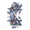
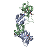
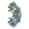
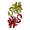
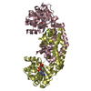
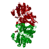
| Title | Crystal structure of the human GIPr ECD in complex with Gipg013 Fab at 3-A resolution | ||||||
 Components Components |
| ||||||
 Keywords Keywords | IMMUNE SYSTEM / Glucagon receptor sub-family recognition fold | ||||||
| Function / homology |  Function and homology information Function and homology informationgastric inhibitory peptide receptor activity / glucagon family peptide binding / gastric inhibitory peptide signaling pathway / endocrine pancreas development / response to fatty acid / desensitization of G protein-coupled receptor signaling pathway / G protein-coupled peptide receptor activity / regulation of insulin secretion / peptide hormone binding / response to axon injury ...gastric inhibitory peptide receptor activity / glucagon family peptide binding / gastric inhibitory peptide signaling pathway / endocrine pancreas development / response to fatty acid / desensitization of G protein-coupled receptor signaling pathway / G protein-coupled peptide receptor activity / regulation of insulin secretion / peptide hormone binding / response to axon injury / response to glucose / activation of adenylate cyclase activity / response to nutrient / generation of precursor metabolites and energy / response to calcium ion / adenylate cyclase-modulating G protein-coupled receptor signaling pathway / positive regulation of insulin secretion / Glucagon-type ligand receptors / transmembrane signaling receptor activity / adenylate cyclase-activating G protein-coupled receptor signaling pathway / positive regulation of cytosolic calcium ion concentration / G alpha (s) signalling events / cell surface receptor signaling pathway / membrane / plasma membrane Similarity search - Function | ||||||
| Biological species |  Homo sapiens (human) Homo sapiens (human) | ||||||
| Method |  X-RAY DIFFRACTION / MOLECULAR REPLACEMENT / Resolution: 3 Å X-RAY DIFFRACTION / MOLECULAR REPLACEMENT / Resolution: 3 Å | ||||||
 Authors Authors | Madhurantakam, C. / Ravn, P. / Gruetter, M.G. / Jackson, R.H. | ||||||
 Citation Citation | Journal: J.Biol.Chem. / Year: 2013 Title: Structural and Pharmacological Characterization of Novel Potent and Selective Monoclonal Antibody Antagonists of Glucose-dependent Insulinotropic Polypeptide Receptor. Authors: Ravn, P. / Madhurantakam, C. / Kunze, S. / Matthews, E. / Priest, C. / O'Brien, S. / Collinson, A. / Papworth, M. / Fritsch-Fredin, M. / Jermutus, L. / Benthem, L. / Gruetter, M. / Jackson, R.H. | ||||||
| History |
|
- Structure visualization
| Structure viewer | Molecule: MolmilJmol/JSmol |
|---|
- Downloads & links
-Download
| PDBx/mmCIF format | 4hj0.cif.gz | 199.6 KB | Display | PDBx/mmCIF format |
|---|---|---|---|---|
| PDB format | pdb4hj0.ent.gz | 158.8 KB | Display | PDB format |
| PDBx/mmJSON format | 4hj0.json.gz | Tree view | PDBx/mmJSON format | |
| Others |  Other downloads Other downloads |
-Validation report
| Arichive directory | https://data.pdbj.org/pub/pdb/validation_reports/hj/4hj0ftp://data.pdbj.org/pub/pdb/validation_reports/hj/4hj0 | HTTPS FTP |
|---|
-Related structure data


-Links
 PDBj
PDBj

- Assembly
| Deposited unit | 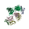
| ||||||||
|---|---|---|---|---|---|---|---|---|---|
| 1 | 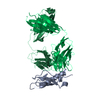
| ||||||||
| 2 | 
| ||||||||
| Unit cell |
|
-Components
| #1: Protein | Mass: 15764.310 Da / Num. of mol.: 2 / Fragment: Extra-cellular Domain, UNP residues 24-138 Source method: isolated from a genetically manipulated source Source: (gene. exp.) Homo sapiens (human) / Gene: GIPR / Plasmid: pET-28a / Production host:  #2: Protein | Mass: 23758.564 Da / Num. of mol.: 2 Source method: isolated from a genetically manipulated source Source: (gene. exp.) Homo sapiens (human) / Plasmid: pEU / Cell line (production host): CHO / Production host: Homo sapiens (human)#3: Protein | Mass: 22691.947 Da / Num. of mol.: 2 Source method: isolated from a genetically manipulated source Source: (gene. exp.) Homo sapiens (human) / Plasmid: pEU / Cell line (production host): CHO / Production host: Homo sapiens (human)Has protein modification | Y | |
|---|
-Experimental details
-Experiment
| Experiment | Method: X-RAY DIFFRACTION / Number of used crystals: 1 |
|---|
- Sample preparation
| Crystal | Density Matthews: 2.24 Å3/Da / Density % sol: 45 % |
|---|---|
| Crystal grow | Temperature: 298 K / Method: vapor diffusion, sitting drop / pH: 9 Details: 0.02 M TAPS, 30% (w/v) PEG 10,000, pH 9, VAPOR DIFFUSION, SITTING DROP, temperature 298K |
-Data collection
| Diffraction | Mean temperature: 100 K |
|---|---|
| Diffraction source | Source: ROTATING ANODE / Type: BRUKER AXS MICROSTAR / Wavelength: 1.54 Å |
| Detector | Type: MAR scanner 345 mm plate / Detector: IMAGE PLATE / Date: Aug 29, 2010 |
| Radiation | Monochromator: Si (III) / Protocol: SINGLE WAVELENGTH / Monochromatic (M) / Laue (L): M / Scattering type: x-ray |
| Radiation wavelength | Wavelength: 1.54 Å / Relative weight: 1 |
| Reflection | Resolution: 3→48.665 Å / Num. obs: 21995 / % possible obs: 100 % / Observed criterion σ(I): 2.6 |
| Reflection shell | Resolution: 3→3.16 Å / % possible all: 100 |
- Processing
| Software |
| |||||||||||||||||||||||||||||||||||||||||||||||||||||||||||||||||||||||||||||||||||||||||||||||||||||||||
|---|---|---|---|---|---|---|---|---|---|---|---|---|---|---|---|---|---|---|---|---|---|---|---|---|---|---|---|---|---|---|---|---|---|---|---|---|---|---|---|---|---|---|---|---|---|---|---|---|---|---|---|---|---|---|---|---|---|---|---|---|---|---|---|---|---|---|---|---|---|---|---|---|---|---|---|---|---|---|---|---|---|---|---|---|---|---|---|---|---|---|---|---|---|---|---|---|---|---|---|---|---|---|---|---|---|---|
| Refinement | Method to determine structure: MOLECULAR REPLACEMENT Starting model: PDB ENTRY 2QKH, 1GIG Resolution: 3→48.665 Å / SU ML: 1 / σ(F): 1.29 / Phase error: 31.06 / Stereochemistry target values: ML
| |||||||||||||||||||||||||||||||||||||||||||||||||||||||||||||||||||||||||||||||||||||||||||||||||||||||||
| Solvent computation | Shrinkage radii: 0.83 Å / VDW probe radii: 1.1 Å / Solvent model: FLAT BULK SOLVENT MODEL / Bsol: 22.75 Å2 / ksol: 0.306 e/Å3 | |||||||||||||||||||||||||||||||||||||||||||||||||||||||||||||||||||||||||||||||||||||||||||||||||||||||||
| Displacement parameters |
| |||||||||||||||||||||||||||||||||||||||||||||||||||||||||||||||||||||||||||||||||||||||||||||||||||||||||
| Refinement step | Cycle: LAST / Resolution: 3→48.665 Å
| |||||||||||||||||||||||||||||||||||||||||||||||||||||||||||||||||||||||||||||||||||||||||||||||||||||||||
| Refine LS restraints |
| |||||||||||||||||||||||||||||||||||||||||||||||||||||||||||||||||||||||||||||||||||||||||||||||||||||||||
| LS refinement shell |
|
