 Movie
Movie Controller

[English] 日本語
 Yorodumi
- PDB-6jx2: Crystal structure of Ketol-acid reductoisomerase from Corynebacte... -
+ Open data

- Basic information
| Entry | Database: PDB / ID: 6jx2 | ||||||
|---|---|---|---|---|---|---|---|
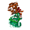
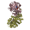
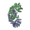
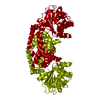
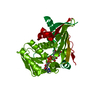
| Title | Crystal structure of Ketol-acid reductoisomerase from Corynebacterium glutamicum | ||||||
 Components Components | Ketol-acid reductoisomerase (NADP(+)) | ||||||
 Keywords Keywords | OXIDOREDUCTASE / Ketol-acid reductoisomerase | ||||||
| Function / homology |  Function and homology information Function and homology information2-dehydropantoate 2-reductase activity / ketol-acid reductoisomerase (NADP+) / ketol-acid reductoisomerase activity / L-valine biosynthetic process / : / NADP binding / magnesium ion binding Similarity search - Function | ||||||
| Biological species |  Corynebacterium glutamicum ATCC 13032 (bacteria) Corynebacterium glutamicum ATCC 13032 (bacteria) | ||||||
| Method |  X-RAY DIFFRACTION / SYNCHROTRON / MOLECULAR REPLACEMENT / Resolution: 2.6 Å X-RAY DIFFRACTION / SYNCHROTRON / MOLECULAR REPLACEMENT / Resolution: 2.6 Å | ||||||
 Authors Authors | Lee, D. / Hong, J. / Kim, K.-J. | ||||||
 Citation Citation | Journal: J.Agric.Food Chem. / Year: 2019 Title: Crystal Structure and Biochemical Characterization of Ketol-Acid Reductoisomerase fromCorynebacterium glutamicum. Authors: Lee, D. / Hong, J. / Kim, K.J. | ||||||
| History |
|
- Structure visualization
| Structure viewer | Molecule: MolmilJmol/JSmol |
|---|
- Downloads & links
-Download
| PDBx/mmCIF format | 6jx2.cif.gz | 255.1 KB | Display | PDBx/mmCIF format |
|---|---|---|---|---|
| PDB format | pdb6jx2.ent.gz | 205 KB | Display | PDB format |
| PDBx/mmJSON format | 6jx2.json.gz | Tree view | PDBx/mmJSON format | |
| Others |  Other downloads Other downloads |
-Validation report
| Arichive directory | https://data.pdbj.org/pub/pdb/validation_reports/jx/6jx2ftp://data.pdbj.org/pub/pdb/validation_reports/jx/6jx2 | HTTPS FTP |
|---|
-Related structure data
| Related structure data |  4ypoS S: Starting model for refinement |
|---|---|
| Similar structure data |


-Links
 PDBj
PDBj


- Assembly
| Deposited unit | 
| ||||||||
|---|---|---|---|---|---|---|---|---|---|
| 1 | 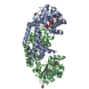
| ||||||||
| 2 | 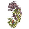
| ||||||||
| Unit cell |
|
-Components
| #1: Protein | Mass: 37021.539 Da / Num. of mol.: 4 Source method: isolated from a genetically manipulated source Source: (gene. exp.) Corynebacterium glutamicum ATCC 13032 (bacteria)Strain: ATCC 13032 / Gene: ilvC, Cgl1273, cg1437 / Plasmid: pET30a / Production host: References: UniProt: Q57179, ketol-acid reductoisomerase (NADP+) #2: Chemical | ChemComp-MG /   Mass: 24.305 Da / Num. of mol.: 13 / Source method: isolated from a natural source / Formula: Mg Mass: 24.305 Da / Num. of mol.: 13 / Source method: isolated from a natural source / Formula: Mg#3: Chemical |   Mass: 743.405 Da / Num. of mol.: 3 / Source method: obtained synthetically / Formula: C21H28N7O17P3 Mass: 743.405 Da / Num. of mol.: 3 / Source method: obtained synthetically / Formula: C21H28N7O17P3#4: Chemical | ChemComp-EDO /   Mass: 62.068 Da / Num. of mol.: 5 / Source method: obtained synthetically / Formula: C2H6O2 Mass: 62.068 Da / Num. of mol.: 5 / Source method: obtained synthetically / Formula: C2H6O2#5: Water | ChemComp-HOH / |  Mass: 18.015 Da / Num. of mol.: 98 / Source method: isolated from a natural source / Formula: H2O Mass: 18.015 Da / Num. of mol.: 98 / Source method: isolated from a natural source / Formula: H2O |
|---|
-Experimental details
-Experiment
| Experiment | Method: X-RAY DIFFRACTION / Number of used crystals: 1 |
|---|
- Sample preparation
| Crystal | Density Matthews: 2.18 Å3/Da / Density % sol: 43.54 % |
|---|---|
| Crystal grow | Temperature: 293 K / Method: vapor diffusion, hanging drop / pH: 5.5 Details: 25 % (w/v) polyethylene glycol (PEG) 3350, 0.1 M Bis-Tris, pH 5.5, 0.2 M Magnesium Chloride |
-Data collection
| Diffraction | Mean temperature: 100 K / Serial crystal experiment: N |
|---|---|
| Diffraction source | Source: SYNCHROTRON / Site: PAL/PLS  / Beamline: 7A (6B, 6C1) / Wavelength: 0.97934 Å / Beamline: 7A (6B, 6C1) / Wavelength: 0.97934 Å |
| Detector | Type: ADSC QUANTUM 270 / Detector: CCD / Date: Nov 6, 2016 |
| Radiation | Monochromator: Double Crystal Monochromator / Protocol: SINGLE WAVELENGTH / Monochromatic (M) / Laue (L): M / Scattering type: x-ray |
| Radiation wavelength | Wavelength: 0.97934 Å / Relative weight: 1 |
| Reflection | Resolution: 2.6→78.91 Å / Num. obs: 37218 / % possible obs: 98.1 % / Redundancy: 4.2 % / Rmerge(I) obs: 0.091 / Net I/σ(I): 20.6 |
| Reflection shell | Resolution: 2.6→2.64 Å / Num. unique obs: 37218 |
- Processing
| Software |
| ||||||||||||||||||||||||||||||||||||||||||||||||||||||||||||
|---|---|---|---|---|---|---|---|---|---|---|---|---|---|---|---|---|---|---|---|---|---|---|---|---|---|---|---|---|---|---|---|---|---|---|---|---|---|---|---|---|---|---|---|---|---|---|---|---|---|---|---|---|---|---|---|---|---|---|---|---|---|
| Refinement | Method to determine structure: MOLECULAR REPLACEMENT Starting model: 4YPO Resolution: 2.6→78.91 Å / Cor.coef. Fo:Fc: 0.951 / Cor.coef. Fo:Fc free: 0.899 / SU B: 14.245 / SU ML: 0.297 / Cross valid method: THROUGHOUT / σ(F): 0 / ESU R Free: 0.378 Details: HYDROGENS HAVE BEEN ADDED IN THE RIDING POSITIONS U VALUES : REFINED INDIVIDUALLY
| ||||||||||||||||||||||||||||||||||||||||||||||||||||||||||||
| Solvent computation | Ion probe radii: 0.8 Å / Shrinkage radii: 0.8 Å / VDW probe radii: 1.2 Å | ||||||||||||||||||||||||||||||||||||||||||||||||||||||||||||
| Displacement parameters | Biso max: 140.24 Å2 / Biso mean: 54.779 Å2 / Biso min: 24.02 Å2
| ||||||||||||||||||||||||||||||||||||||||||||||||||||||||||||
| Refinement step | Cycle: final / Resolution: 2.6→78.91 Å
| ||||||||||||||||||||||||||||||||||||||||||||||||||||||||||||
| Refine LS restraints |
| ||||||||||||||||||||||||||||||||||||||||||||||||||||||||||||
| LS refinement shell | Resolution: 2.601→2.669 Å / Rfactor Rfree error: 0 / Total num. of bins used: 20
|
