 Movie
Movie Controller

[English] 日本語
 Yorodumi
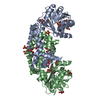
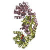
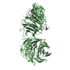
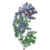
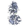
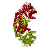
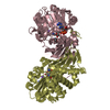
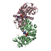
- PDB-6l2s: IlvC, a ketol-acid reductoisomerase, from Streptococcus pneumonia... -
+ Open data

- Basic information
| Entry | Database: PDB / ID: 6l2s | |||||||||||||||
|---|---|---|---|---|---|---|---|---|---|---|---|---|---|---|---|---|
| Title | IlvC, a ketol-acid reductoisomerase, from Streptococcus pneumoniae_D83G | |||||||||||||||
 Components Components | Ketol-acid reductoisomerase (NADP(+)) | |||||||||||||||
 Keywords Keywords | ISOMERASE / IlvC / Stereptococcus pneumoniae / BCAA pathway / ketol-acid reductoisomerase / D83G | |||||||||||||||
| Function / homology |  Function and homology information Function and homology informationketol-acid reductoisomerase (NADP+) / ketol-acid reductoisomerase activity / L-valine biosynthetic process / : / NADP binding / magnesium ion binding / cytosol Similarity search - Function | |||||||||||||||
| Biological species |  Streptococcus pneumoniae D39 (bacteria) Streptococcus pneumoniae D39 (bacteria) | |||||||||||||||
| Method |  X-RAY DIFFRACTION / SYNCHROTRON / MOLECULAR REPLACEMENT / Resolution: 2.29 Å X-RAY DIFFRACTION / SYNCHROTRON / MOLECULAR REPLACEMENT / Resolution: 2.29 Å | |||||||||||||||
 Authors Authors | Gyuhee, K. / Donghyuk, S. / Sumin, L. / Jaesook, Y. / Sangho, L. | |||||||||||||||
| Funding support |  Korea, Republic Of, 4items Korea, Republic Of, 4items
| |||||||||||||||
 Citation Citation | Journal: Crystals / Year: 2019 Title: Crystal Structure of IlvC, a Ketol-Acid Reductoisomerase, from Streptococcus Pneumoniae. Authors: Kim, G.H. / Shin, D.H. / Lee, S.M. / Yoon, J.S. / Lee, S.H. | |||||||||||||||
| History |
|
- Structure visualization
| Structure viewer | Molecule: MolmilJmol/JSmol |
|---|
- Downloads & links
-Download
| PDBx/mmCIF format | 6l2s.cif.gz | 161.7 KB | Display | PDBx/mmCIF format |
|---|---|---|---|---|
| PDB format | pdb6l2s.ent.gz | 103.4 KB | Display | PDB format |
| PDBx/mmJSON format | 6l2s.json.gz | Tree view | PDBx/mmJSON format | |
| Others |  Other downloads Other downloads |
-Validation report
| Arichive directory | https://data.pdbj.org/pub/pdb/validation_reports/l2/6l2sftp://data.pdbj.org/pub/pdb/validation_reports/l2/6l2s | HTTPS FTP |
|---|
-Related structure data
| Related structure data |  6l2iSC 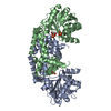 6l2kC 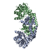 6l2rC 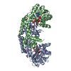 6l2zC S: Starting model for refinement C: citing same article ( |
|---|---|
| Similar structure data |


-Links
 PDBj
PDBj


- Assembly
| Deposited unit | 
| ||||||||||||
|---|---|---|---|---|---|---|---|---|---|---|---|---|---|
| 1 |
| ||||||||||||
| Unit cell |
|
-Components
| #1: Protein | Mass: 37334.195 Da / Num. of mol.: 2 / Mutation: D83G Source method: isolated from a genetically manipulated source Source: (gene. exp.) Streptococcus pneumoniae D39 (bacteria)Strain: D39 / NCTC 7466 / Gene: ilvC, SPD_0406 / Production host: References: UniProt: Q04M32, ketol-acid reductoisomerase (NADP+) Has protein modification | Y | |
|---|
-Experimental details
-Experiment
| Experiment | Method: X-RAY DIFFRACTION / Number of used crystals: 1 |
|---|
- Sample preparation
| Crystal | Density Matthews: 2.74 Å3/Da / Density % sol: 55.03 % |
|---|---|
| Crystal grow | Temperature: 295 K / Method: vapor diffusion, hanging drop Details: 0.1 M HEPES pH 7.5, 0.1 M NaCl, 1.8 M ammonium sulfate |
-Data collection
| Diffraction | Mean temperature: 100 K / Serial crystal experiment: N |
|---|---|
| Diffraction source | Source: SYNCHROTRON / Site: PAL/PLS / Beamline: 7A (6B, 6C1) / Wavelength: 1 Å |
| Detector | Type: ADSC QUANTUM 270 / Detector: CCD / Date: Oct 11, 2014 |
| Radiation | Protocol: SINGLE WAVELENGTH / Monochromatic (M) / Laue (L): M / Scattering type: x-ray |
| Radiation wavelength | Wavelength: 1 Å / Relative weight: 1 |
| Reflection | Resolution: 2.29→30.3 Å / Num. obs: 35335 / % possible obs: 99.8 % / Redundancy: 7.1 % / Biso Wilson estimate: 28.3 Å2 / CC1/2: 0.997 / Net I/σ(I): 50 |
| Reflection shell | Resolution: 2.29→2.37 Å / Num. unique obs: 1753 / CC1/2: 0.972 |
- Processing
| Software |
| ||||||||||||||||||||||||||||||||||||||||||||||||||||||||||||||||||||||||||||||||||||||||||||||||||
|---|---|---|---|---|---|---|---|---|---|---|---|---|---|---|---|---|---|---|---|---|---|---|---|---|---|---|---|---|---|---|---|---|---|---|---|---|---|---|---|---|---|---|---|---|---|---|---|---|---|---|---|---|---|---|---|---|---|---|---|---|---|---|---|---|---|---|---|---|---|---|---|---|---|---|---|---|---|---|---|---|---|---|---|---|---|---|---|---|---|---|---|---|---|---|---|---|---|---|---|
| Refinement | Method to determine structure: MOLECULAR REPLACEMENT Starting model: 6L2I Resolution: 2.29→30.3 Å / SU ML: 0.2605 / Cross valid method: FREE R-VALUE / σ(F): 1.36 / Phase error: 24.1942
| ||||||||||||||||||||||||||||||||||||||||||||||||||||||||||||||||||||||||||||||||||||||||||||||||||
| Solvent computation | Shrinkage radii: 0.9 Å / VDW probe radii: 1.11 Å | ||||||||||||||||||||||||||||||||||||||||||||||||||||||||||||||||||||||||||||||||||||||||||||||||||
| Displacement parameters | Biso mean: 32.3 Å2 | ||||||||||||||||||||||||||||||||||||||||||||||||||||||||||||||||||||||||||||||||||||||||||||||||||
| Refinement step | Cycle: LAST / Resolution: 2.29→30.3 Å
| ||||||||||||||||||||||||||||||||||||||||||||||||||||||||||||||||||||||||||||||||||||||||||||||||||
| Refine LS restraints |
| ||||||||||||||||||||||||||||||||||||||||||||||||||||||||||||||||||||||||||||||||||||||||||||||||||
| LS refinement shell |
|
