 Movie
Movie Controller

[English] 日本語
 Yorodumi
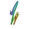
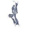
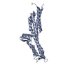
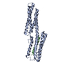
- PDB-3k6b: X-ray crystal structure of the E2 domain of APL-1 from C. elegans... -
+ Open data

- Basic information
| Entry | Database: PDB / ID: 3k6b | |||||||||
|---|---|---|---|---|---|---|---|---|---|---|
| Title | X-ray crystal structure of the E2 domain of APL-1 from C. elegans, in complex with sucrose octasulfate (SOS) | |||||||||
 Components Components | Beta-amyloid-like protein | |||||||||
 Keywords Keywords | CELL ADHESION / X-ray crystal structure / amyloid precursor protein / heparin binding / Alternative splicing / Amyloid / Developmental protein / Differentiation / Glycoprotein / Membrane / Neurogenesis / Transmembrane | |||||||||
| Function / homology |  Function and homology information Function and homology informationG alpha (q) signalling events / Insertion of tail-anchored proteins into the endoplasmic reticulum membrane / Regulation of Insulin-like Growth Factor (IGF) transport and uptake by Insulin-like Growth Factor Binding Proteins (IGFBPs) / Post-translational protein phosphorylation / ecdysis, collagen and cuticulin-based cuticle / Platelet degranulation / ECM proteoglycans / body morphogenesis / nematode larval development / transition metal ion binding ...G alpha (q) signalling events / Insertion of tail-anchored proteins into the endoplasmic reticulum membrane / Regulation of Insulin-like Growth Factor (IGF) transport and uptake by Insulin-like Growth Factor Binding Proteins (IGFBPs) / Post-translational protein phosphorylation / ecdysis, collagen and cuticulin-based cuticle / Platelet degranulation / ECM proteoglycans / body morphogenesis / nematode larval development / transition metal ion binding / axonogenesis / central nervous system development / heparin binding / cytoplasmic vesicle / early endosome / neuron projection / neuronal cell body / membrane Similarity search - Function | |||||||||
| Biological species |  | |||||||||
| Method |  X-RAY DIFFRACTION / SYNCHROTRON / MOLECULAR REPLACEMENT / Resolution: 2.8 Å X-RAY DIFFRACTION / SYNCHROTRON / MOLECULAR REPLACEMENT / Resolution: 2.8 Å | |||||||||
 Authors Authors | Hoopes, J.T. / Ha, Y. | |||||||||
 Citation Citation | Journal: J.Biol.Chem. / Year: 2010 Title: Structural characterization of the E2 domain of APL-1, a Caenorhabditis elegans homolog of human amyloid precursor protein, and its heparin binding site Authors: Hoopes, J.T. / Liu, X. / Xu, X. / Demeler, B. / Folta-Stogniew, E. / Li, C. / Ha, Y. #1: Journal: Mol.Cell / Year: 2004Title: The X-ray structure of an antiparallel dimer of the human amyloid precursor protein E2 domain. Authors: Wang, Y. / Ha, Y. | |||||||||
| History |
|
- Structure visualization
| Structure viewer | Molecule: MolmilJmol/JSmol |
|---|
- Downloads & links
-Download
| PDBx/mmCIF format | 3k6b.cif.gz | 58.2 KB | Display | PDBx/mmCIF format |
|---|---|---|---|---|
| PDB format | pdb3k6b.ent.gz | 41.3 KB | Display | PDB format |
| PDBx/mmJSON format | 3k6b.json.gz | Tree view | PDBx/mmJSON format | |
| Others |  Other downloads Other downloads |
-Validation report
| Arichive directory | https://data.pdbj.org/pub/pdb/validation_reports/k6/3k6bftp://data.pdbj.org/pub/pdb/validation_reports/k6/3k6b | HTTPS FTP |
|---|
-Related structure data
| Related structure data |  3k66SC S: Starting model for refinement C: citing same article ( |
|---|---|
| Similar structure data |


-Links
 PDBj
PDBj

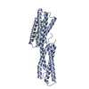
- Assembly
| Deposited unit | 
| ||||||||
|---|---|---|---|---|---|---|---|---|---|
| 1 |
| ||||||||
| Unit cell |
|
-Components
| #1: Protein | Mass: 28516.445 Da / Num. of mol.: 1 / Fragment: E2 Domain: UNP residues 240-478' Source method: isolated from a genetically manipulated source Source: (gene. exp.)  |
|---|---|
| #2: Sugar | ChemComp-GU4 / 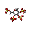  Type: D-saccharide, alpha linking / Mass: 500.409 Da / Num. of mol.: 1 Type: D-saccharide, alpha linking / Mass: 500.409 Da / Num. of mol.: 1Source method: isolated from a genetically manipulated source Formula: C6H12O18S4 |
| #3: Water | ChemComp-HOH /  Mass: 18.015 Da / Num. of mol.: 34 / Source method: isolated from a natural source / Formula: H2O Mass: 18.015 Da / Num. of mol.: 34 / Source method: isolated from a natural source / Formula: H2O |
-Experimental details
-Experiment
| Experiment | Method: X-RAY DIFFRACTION / Number of used crystals: 1 |
|---|
- Sample preparation
| Crystal | Density Matthews: 3.23 Å3/Da / Density % sol: 61.96 % |
|---|---|
| Crystal grow | Temperature: 298 K / Method: vapor diffusion, hanging drop / pH: 7 Details: 20% PEG 3350, 15% Isopropanol, 0.1 M Hepes pH 7.0, VAPOR DIFFUSION, HANGING DROP, temperature 298K |
-Data collection
| Diffraction | Mean temperature: 100 K |
|---|---|
| Diffraction source | Source: SYNCHROTRON / Site: NSLS  / Beamline: X29A / Wavelength: 1.0809 Å / Beamline: X29A / Wavelength: 1.0809 Å |
| Detector | Type: ADSC QUANTUM 210 / Detector: CCD / Date: May 20, 2007 |
| Radiation | Protocol: SINGLE WAVELENGTH / Monochromatic (M) / Laue (L): M / Scattering type: x-ray |
| Radiation wavelength | Wavelength: 1.0809 Å / Relative weight: 1 |
| Reflection | Resolution: 2.8→40 Å / Num. obs: 9028 / Redundancy: 9.6 % / Rsym value: 0.053 / Net I/σ(I): 20.7 |
| Reflection shell | Resolution: 2.8→2.9 Å / Mean I/σ(I) obs: 2.6 / Rsym value: 0.346 |
- Processing
| Software |
| ||||||||||||||||
|---|---|---|---|---|---|---|---|---|---|---|---|---|---|---|---|---|---|
| Refinement | Method to determine structure: MOLECULAR REPLACEMENT Starting model: PDB entry 3K66 Resolution: 2.8→40 Å / σ(F): 2.6
| ||||||||||||||||
| Displacement parameters | Biso mean: 84.5 Å2 | ||||||||||||||||
| Refinement step | Cycle: LAST / Resolution: 2.8→40 Å
| ||||||||||||||||
| Refine LS restraints |
|
