 Movie
Movie Controller

[English] 日本語
 Yorodumi
- PDB-3jxp: Crystal Structure of the Coenzyme PQQ Synthesis Protein (PqqB) fr... -
+ Open data

- Basic information
| Entry | Database: PDB / ID: 3jxp | ||||||
|---|---|---|---|---|---|---|---|
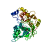
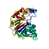
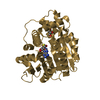
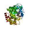
| Title | Crystal Structure of the Coenzyme PQQ Synthesis Protein (PqqB) from Pseudomonas putida | ||||||
 Components Components | Coenzyme PQQ synthesis protein B | ||||||
 Keywords Keywords | BIOSYNTHETIC PROTEIN / alpha-beta protein / pqq biosynthesis / Transport | ||||||
| Function / homology |  Function and homology information Function and homology information | ||||||
| Biological species |  Pseudomonas putida (bacteria) Pseudomonas putida (bacteria) | ||||||
| Method |  X-RAY DIFFRACTION / SYNCHROTRON / MOLECULAR REPLACEMENT / Resolution: 2.2 Å X-RAY DIFFRACTION / SYNCHROTRON / MOLECULAR REPLACEMENT / Resolution: 2.2 Å | ||||||
 Authors Authors | Metlitzky, M. / Schwarzenbacher, R. | ||||||
 Citation Citation | Journal: J. Biophys. Chem. / Year: 2012 Title: Crystal structure of PqqB from Pseudomonas putida at 2.2 A resolution Authors: Metlitzky, M. / Puehringer, S. / Fisher, S.J. | ||||||
| History |
|
- Structure visualization
| Structure viewer | Molecule: MolmilJmol/JSmol |
|---|
- Downloads & links
-Download
| PDBx/mmCIF format | 3jxp.cif.gz | 77.3 KB | Display | PDBx/mmCIF format |
|---|---|---|---|---|
| PDB format | pdb3jxp.ent.gz | 56.2 KB | Display | PDB format |
| PDBx/mmJSON format | 3jxp.json.gz | Tree view | PDBx/mmJSON format | |
| Others |  Other downloads Other downloads |
-Validation report
| Arichive directory | https://data.pdbj.org/pub/pdb/validation_reports/jx/3jxpftp://data.pdbj.org/pub/pdb/validation_reports/jx/3jxp | HTTPS FTP |
|---|
-Related structure data
| Related structure data | 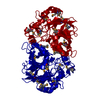 1xtoS S: Starting model for refinement |
|---|---|
| Similar structure data |


-Links
 PDBj
PDBj

- Assembly
| Deposited unit | 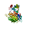
| ||||||||
|---|---|---|---|---|---|---|---|---|---|
| 1 |
| ||||||||
| 2 | 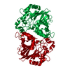
| ||||||||
| Unit cell |
| ||||||||
| Details | monomer |
-Components
| #1: Protein | Mass: 35740.426 Da / Num. of mol.: 1 / Mutation: M253T Source method: isolated from a genetically manipulated source Source: (gene. exp.) Pseudomonas putida (bacteria) / Strain: KT2440 / Gene: PP_0379, pqqB / Plasmid: pET21 / Production host: | ||
|---|---|---|---|
| #2: Chemical | ChemComp-ZN /   Mass: 65.409 Da / Num. of mol.: 1 / Source method: obtained synthetically / Formula: Zn Mass: 65.409 Da / Num. of mol.: 1 / Source method: obtained synthetically / Formula: Zn | ||
| #3: Chemical |   Mass: 35.453 Da / Num. of mol.: 2 / Source method: obtained synthetically / Formula: Cl Mass: 35.453 Da / Num. of mol.: 2 / Source method: obtained synthetically / Formula: Cl#4: Water | ChemComp-HOH / |  Mass: 18.015 Da / Num. of mol.: 174 / Source method: isolated from a natural source / Formula: H2O Mass: 18.015 Da / Num. of mol.: 174 / Source method: isolated from a natural source / Formula: H2O |
-Experimental details
-Experiment
| Experiment | Method: X-RAY DIFFRACTION / Number of used crystals: 1 |
|---|
- Sample preparation
| Crystal | Density Matthews: 2.84 Å3/Da / Density % sol: 56.75 % |
|---|---|
| Crystal grow | Temperature: 293 K / Method: vapor diffusion, sitting drop / pH: 8.5 Details: 0.1 M TRIS hydrochloride pH 8.5, 0.2 M Magnesium chloride hexahydrate, 15% w/v PEG 4,000, VAPOR DIFFUSION, SITTING DROP, temperature 293K |
-Data collection
| Diffraction | Mean temperature: 100 K |
|---|---|
| Diffraction source | Source: SYNCHROTRON / Site: BESSY  / Beamline: 14.1 / Wavelength: 0.91841 Å / Beamline: 14.1 / Wavelength: 0.91841 Å |
| Detector | Type: RAYONIX MX-225 / Detector: CCD / Date: Apr 18, 2009 |
| Radiation | Monochromator: Double crystal, Si(111) / Protocol: SINGLE WAVELENGTH / Monochromatic (M) / Laue (L): M / Scattering type: x-ray |
| Radiation wavelength | Wavelength: 0.91841 Å / Relative weight: 1 |
| Reflection | Resolution: 2.2→67.7 Å / Num. all: 21579 / Num. obs: 21411 / % possible obs: 98.2 % |
| Reflection shell | Resolution: 2.2→2.257 Å / Rmerge(I) obs: 0.569 / % possible all: 99.62 |
- Processing
| Software |
| ||||||||||||||||||||||||||||||||||||||||||||||||||||||||||||||||||||||||||||||||||||||||||
|---|---|---|---|---|---|---|---|---|---|---|---|---|---|---|---|---|---|---|---|---|---|---|---|---|---|---|---|---|---|---|---|---|---|---|---|---|---|---|---|---|---|---|---|---|---|---|---|---|---|---|---|---|---|---|---|---|---|---|---|---|---|---|---|---|---|---|---|---|---|---|---|---|---|---|---|---|---|---|---|---|---|---|---|---|---|---|---|---|---|---|---|
| Refinement | Method to determine structure: MOLECULAR REPLACEMENT Starting model: PDB ENTRY 1XTO Resolution: 2.2→67.7 Å / Cor.coef. Fo:Fc: 0.942 / Cor.coef. Fo:Fc free: 0.916 / SU B: 4.926 / SU ML: 0.129 / Cross valid method: THROUGHOUT / ESU R: 0.22 / ESU R Free: 0.193 / Stereochemistry target values: MAXIMUM LIKELIHOOD / Details: HYDROGENS HAVE BEEN ADDED IN THE RIDING POSITIONS
| ||||||||||||||||||||||||||||||||||||||||||||||||||||||||||||||||||||||||||||||||||||||||||
| Solvent computation | Ion probe radii: 0.8 Å / Shrinkage radii: 0.8 Å / VDW probe radii: 1.2 Å / Solvent model: MASK | ||||||||||||||||||||||||||||||||||||||||||||||||||||||||||||||||||||||||||||||||||||||||||
| Displacement parameters | Biso mean: 29.329 Å2
| ||||||||||||||||||||||||||||||||||||||||||||||||||||||||||||||||||||||||||||||||||||||||||
| Refinement step | Cycle: LAST / Resolution: 2.2→67.7 Å
| ||||||||||||||||||||||||||||||||||||||||||||||||||||||||||||||||||||||||||||||||||||||||||
| Refine LS restraints |
| ||||||||||||||||||||||||||||||||||||||||||||||||||||||||||||||||||||||||||||||||||||||||||
| LS refinement shell | Resolution: 2.2→2.257 Å / Total num. of bins used: 20
|
