ムービー
ムービー コントローラー


+ データを開く

- 基本情報
基本情報
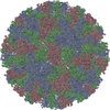
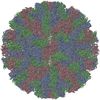
| 登録情報 | データベース: PDB / ID: 3j24 | ||||||
|---|---|---|---|---|---|---|---|
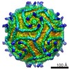
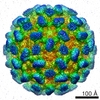
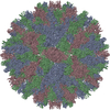
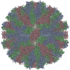
| タイトル | CryoEM reconstruction of complement decay-accelerating factor | ||||||
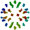
 要素 要素 | Complement decay-accelerating factor | ||||||
 キーワード キーワード | IMMUNE SYSTEM / blood group antigen / complement pathway / glycoprotein / GPI-anchor / immune response / innate immunity / lipoprotein / membrane / Sushi | ||||||
| 機能・相同性 |  機能・相同性情報 機能・相同性情報regulation of lipopolysaccharide-mediated signaling pathway / negative regulation of complement activation / regulation of complement-dependent cytotoxicity / regulation of complement activation / respiratory burst / positive regulation of CD4-positive, alpha-beta T cell activation / positive regulation of CD4-positive, alpha-beta T cell proliferation / Class B/2 (Secretin family receptors) / ficolin-1-rich granule membrane / COPI-mediated anterograde transport ...regulation of lipopolysaccharide-mediated signaling pathway / negative regulation of complement activation / regulation of complement-dependent cytotoxicity / regulation of complement activation / respiratory burst / positive regulation of CD4-positive, alpha-beta T cell activation / positive regulation of CD4-positive, alpha-beta T cell proliferation / Class B/2 (Secretin family receptors) / ficolin-1-rich granule membrane / COPI-mediated anterograde transport / side of membrane / complement activation, classical pathway / transport vesicle / endoplasmic reticulum-Golgi intermediate compartment membrane / secretory granule membrane / Regulation of Complement cascade / positive regulation of T cell cytokine production / virus receptor activity / positive regulation of cytosolic calcium ion concentration / membrane raft / Golgi membrane / innate immune response / lipid binding / Neutrophil degranulation / cell surface / extracellular exosome / extracellular region / plasma membrane 類似検索 - 分子機能 | ||||||
| 生物種 |  Homo sapiens (ヒト) Homo sapiens (ヒト) | ||||||
| 手法 | 電子顕微鏡法 / 単粒子再構成法 / クライオ電子顕微鏡法 / 解像度: 9 Å | ||||||
 データ登録者 データ登録者 | Yoder, J.D. / Hafenstein, S.H. | ||||||
 引用 引用 | ジャーナル: J Virol / 年: 2012 タイトル: The crystal structure of a coxsackievirus B3-RD variant and a refined 9-angstrom cryo-electron microscopy reconstruction of the virus complexed with decay-accelerating factor (DAF) ...タイトル: The crystal structure of a coxsackievirus B3-RD variant and a refined 9-angstrom cryo-electron microscopy reconstruction of the virus complexed with decay-accelerating factor (DAF) provide a new footprint of DAF on the virus surface. 著者: Joshua D Yoder / Javier O Cifuente / Jieyan Pan / Jeffrey M Bergelson / Susan Hafenstein /  要旨: The coxsackievirus-adenovirus receptor (CAR) and decay-accelerating factor (DAF) have been identified as cellular receptors for coxsackievirus B3 (CVB3). The first described DAF-binding isolate was ...The coxsackievirus-adenovirus receptor (CAR) and decay-accelerating factor (DAF) have been identified as cellular receptors for coxsackievirus B3 (CVB3). The first described DAF-binding isolate was obtained during passage of the prototype strain, Nancy, on rhabdomyosarcoma (RD) cells, which express DAF but very little CAR. Here, the structure of the resulting variant, CVB3-RD, has been solved by X-ray crystallography to 2.74 Å, and a cryo-electron microscopy reconstruction of CVB3-RD complexed with DAF has been refined to 9.0 Å. This new high-resolution structure permits us to correct an error in our previous view of DAF-virus interactions, providing a new footprint of DAF that bridges two adjacent protomers. The contact sites between the virus and DAF clearly encompass CVB3-RD residues recently shown to be required for binding to DAF; these residues interact with DAF short consensus repeat 2 (SCR2), which is known to be essential for virus binding. Based on the new structure, the mode of the DAF interaction with CVB3 differs significantly from the mode reported previously for DAF binding to echoviruses. | ||||||
| 履歴 |
|
- 構造の表示
構造の表示
| ムービー |
ムービービューア |
|---|---|
| 構造ビューア | 分子: MolmilJmol/JSmol |
 UCSF Chimera
- ダウンロードとリンク
-ダウンロード
| PDBx/mmCIF形式 | 3j24.cif.gz | 57.3 KB | 表示 | PDBx/mmCIF形式 |
|---|---|---|---|---|
| PDB形式 | pdb3j24.ent.gz | 44.6 KB | 表示 | PDB形式 |
| PDBx/mmJSON形式 | 3j24.json.gz | ツリー表示 | PDBx/mmJSON形式 | |
| その他 |  その他のダウンロード その他のダウンロード |
-検証レポート
| 文書・要旨 | 3j24_validation.pdf.gz | 785.4 KB | 表示 | wwPDB検証レポート |
|---|---|---|---|---|
| 文書・詳細版 | 3j24_full_validation.pdf.gz | 800.1 KB | 表示 | |
| XML形式データ | 3j24_validation.xml.gz | 16.4 KB | 表示 | |
| CIF形式データ | 3j24_validation.cif.gz | 22.4 KB | 表示 | |
| アーカイブディレクトリ | https://data.pdbj.org/pub/pdb/validation_reports/j2/3j24ftp://data.pdbj.org/pub/pdb/validation_reports/j2/3j24 | HTTPS FTP |
-関連構造データ

-リンク
 PDBj
PDBj


- 集合体
集合体
| 登録構造単位 | 
|
|---|---|
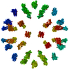
| 1 | x 60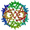
|
| 2 |
|
| 3 | x 5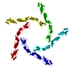
|
| 4 | x 6
|
| 5 | 
|
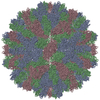
| 対称性 | 点対称性: (シェーンフリース記号: I (正20面体型対称)) |
-要素
| #1: タンパク質 | 分子量: 28174.666 Da / 分子数: 1 断片: four extracellular SCR domains (UNP residues 35-285) 由来タイプ: 組換発現 / 由来: (組換発現) Homo sapiens (ヒト) / 遺伝子: CD55, CR, DAF / 発現宿主:  |
|---|
-実験情報
-実験
| 実験 | 手法: 電子顕微鏡法 |
|---|---|
| EM実験 | 試料の集合状態: PARTICLE / 3次元再構成法: 単粒子再構成法 |
- 試料調製
試料調製
| 構成要素 | 名称: Complement decay-accelerating factor bound to coxsackievirus B3-RD strain タイプ: VIRUS |
|---|---|
| 緩衝液 | 名称: 2-(N-morpholino)ethanesulfonic acid (MES) / pH: 6 / 詳細: 50mM MES |
| 試料 | 濃度: 2 mg/ml / 包埋: NO / シャドウイング: NO / 染色: NO / 凍結: YES |
| 試料支持 | 詳細: Quantifoil |
| 急速凍結 | 装置: HOMEMADE PLUNGER / 凍結剤: ETHANE / Temp: 120 K 詳細: Blotted before plunging in liquid ethane (homemade plunger). |
- 電子顕微鏡撮影
| 顕微鏡 | モデル: FEI/PHILIPS CM300FEG/T / 日付: 2006年8月4日 |
|---|---|
| 電子銃 | 電子線源: TUNGSTEN HAIRPIN / 加速電圧: 300 kV / 照射モード: FLOOD BEAM |
| 電子レンズ | モード: BRIGHT FIELD / 倍率(公称値): 45000 X / 倍率(補正後): 47000 X / 最大 デフォーカス(公称値): 4600 nm / 最小 デフォーカス(公称値): 1000 nm / Cs: 2 mm |
| 試料ホルダ | 試料ホルダーモデル: GATAN LIQUID NITROGEN / 資料ホルダタイプ: Side mounted nitrogen cooled / 温度: 93 K / 最高温度: 93 K / 最低温度: 83 K / 傾斜角・最大: 0 ° / 傾斜角・最小: 0 ° |
| 撮影 | 電子線照射量: 24 e/Å2 / フィルム・検出器のモデル: KODAK SO-163 FILM |
| 画像スキャン | デジタル画像の数: 36 |
| 放射 | プロトコル: SINGLE WAVELENGTH / 単色(M)・ラウエ(L): M / 散乱光タイプ: x-ray |
| 放射波長 | 相対比: 1 |
- 解析
解析
| EMソフトウェア |
| ||||||||||||||||||
|---|---|---|---|---|---|---|---|---|---|---|---|---|---|---|---|---|---|---|---|
| CTF補正 | 詳細: AUTO3DEM | ||||||||||||||||||
| 対称性 | 点対称性: I (正20面体型対称) | ||||||||||||||||||
| 3次元再構成 | 手法: common lines / 解像度: 9 Å / 粒子像の数: 3010 / ピクセルサイズ(公称値): 2.94 Å / ピクセルサイズ(実測値): 2.94 Å / 対称性のタイプ: POINT | ||||||||||||||||||
| 原子モデル構築 | プロトコル: RIGID BODY FIT / 空間: REAL / Target criteria: average map value 詳細: METHOD--Local refinement, rigid body REFINEMENT PROTOCOL--rigid body | ||||||||||||||||||
| 原子モデル構築 | PDB-ID: 1OJW PDB chain-ID: B | ||||||||||||||||||
| 精密化ステップ | サイクル: LAST
|