 Movie
Movie Controller

[English] 日本語
 Yorodumi
- PDB-3j1s: Structure of adeno-associated virus-2 in complex with neutralizin... -
+ Open data

- Basic information
| Entry | Database: PDB / ID: 3j1s | ||||||
|---|---|---|---|---|---|---|---|
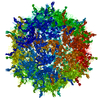
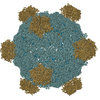
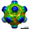
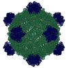
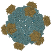
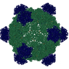
| Title | Structure of adeno-associated virus-2 in complex with neutralizing monoclonal antibody A20 | ||||||
 Components Components |
| ||||||
 Keywords Keywords | VIRUS/IMMUNE SYSTEM / Epitope / Fab / gene therapy / VIRUS-IMMUNE SYSTEM complex | ||||||
| Function / homology |  Function and homology information Function and homology informationsymbiont entry into host cell via permeabilization of host membrane / host cell nucleolus / T=1 icosahedral viral capsid / clathrin-dependent endocytosis of virus by host cell / virion attachment to host cell / structural molecule activity Similarity search - Function | ||||||
| Biological species |   Adeno-associated virus - 2 Adeno-associated virus - 2 | ||||||
| Method | ELECTRON MICROSCOPY / single particle reconstruction / cryo EM / Resolution: 8.5 Å | ||||||
 Authors Authors | Chapman, M.S. / McCraw, D.M. | ||||||
 Citation Citation | Journal: Virology Title: Structure of adeno-associated virus-2 in complex with neutralizing monoclonal antibody A20. Authors: Dustin M McCraw / Jason K O'Donnell / Kenneth A Taylor / Scott M Stagg / Michael S Chapman /  Abstract: The use of adeno-associated virus (AAV) as a gene therapy vector is limited by the host neutralizing immune response. The cryo-electron microscopy (EM) structure at 8.5Å resolution is determined for ...The use of adeno-associated virus (AAV) as a gene therapy vector is limited by the host neutralizing immune response. The cryo-electron microscopy (EM) structure at 8.5Å resolution is determined for a complex of AAV-2 with the Fab' fragment of monoclonal antibody (MAb) A20, the most extensively characterized AAV MAb. The binding footprint is determined through fitting the cryo-EM reconstruction with a homology model following sequencing of the variable domain, and provides a structural basis for integrating diverse prior epitope mappings. The footprint extends from the previously implicated plateau to the side of the spike, and into the conserved canyon, covering a larger area than anticipated. Comparison with structures of binding and non-binding serotypes indicates that recognition depends on a combination of subtle serotype-specific features. Separation of the neutralizing epitope from the heparan sulfate cell attachment site encourages attempts to develop immune-resistant vectors that can still bind to target cells. | ||||||
| History |
|
- Structure visualization
| Movie |
Movie viewer |
|---|---|
| Structure viewer | Molecule: MolmilJmol/JSmol |
- Downloads & links
-Download
| PDBx/mmCIF format | 3j1s.cif.gz | 190.9 KB | Display | PDBx/mmCIF format |
|---|---|---|---|---|
| PDB format | pdb3j1s.ent.gz | 145.6 KB | Display | PDB format |
| PDBx/mmJSON format | 3j1s.json.gz | Tree view | PDBx/mmJSON format | |
| Others |  Other downloads Other downloads |
-Validation report
| Arichive directory | https://data.pdbj.org/pub/pdb/validation_reports/j1/3j1sftp://data.pdbj.org/pub/pdb/validation_reports/j1/3j1s | HTTPS FTP |
|---|
-Related structure data
| Related structure data | 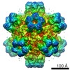 5424MC M: map data used to model this data C: citing same article ( |
|---|---|
| Similar structure data |
-Links
 PDBj
PDBj


- Assembly
| Deposited unit | 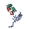
|
|---|---|
| 1 | x 60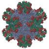
|
| 2 |
|
| 3 | x 5
|
| 4 | x 6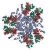
|
| 5 | 
|
| Symmetry | Point symmetry: (Schoenflies symbol: I (icosahedral)) |
-Components
| #1: Antibody | Mass: 23603.965 Da / Num. of mol.: 1 / Fragment: Fab' / Source method: isolated from a natural source / Source: (natural) |
|---|---|
| #2: Antibody | Mass: 23777.314 Da / Num. of mol.: 1 / Fragment: Fab' / Source method: isolated from a natural source / Source: (natural) |
| #3: Protein | Mass: 58772.625 Da / Num. of mol.: 1 / Fragment: SEE REMARK 999 / Source method: isolated from a natural source / Source: (natural) Adeno-associated virus - 2 / References: UniProt: P03135 |
| Has protein modification | Y |
| Sequence details | DUE TO IN-FRAME ALTERNATIVE SPLICING, THE AAV-2 CAPSID PROTEIN IN THIS ENTRY IS A 1:1:10 MIXTURE OF ...DUE TO IN-FRAME ALTERNATIV |
-Experimental details
-Experiment
| Experiment | Method: ELECTRON MICROSCOPY |
|---|---|
| EM experiment | Aggregation state: PARTICLE / 3D reconstruction method: single particle reconstruction |
- Sample preparation
| Component |
| ||||||||||||||||||||||||||||||
|---|---|---|---|---|---|---|---|---|---|---|---|---|---|---|---|---|---|---|---|---|---|---|---|---|---|---|---|---|---|---|---|
| Molecular weight | Value: 6.9 MDa / Experimental value: NO | ||||||||||||||||||||||||||||||
| Details of virus | Details: Single-stranded DNA Parvovirus / Empty: NO / Enveloped: NO / Host category: VERTEBRATES / Isolate: SEROTYPE / Type: VIRION | ||||||||||||||||||||||||||||||
| Natural host | Organism: Homo sapiens / Strain: HeLa | ||||||||||||||||||||||||||||||
| Buffer solution | Name: 100 mM HEPES, 50 mM magnesium chloride, 5% glycerol / pH: 7.2 Details: 100 mM HEPES, 50 mM magnesium chloride, 5% glycerol | ||||||||||||||||||||||||||||||
| Specimen | Conc.: 0.14 mg/ml / Embedding applied: NO / Shadowing applied: NO / Staining applied: NO / Vitrification applied: YES Details: 100 mM HEPES, 50 mM magnesium chloride, 5% glycerol | ||||||||||||||||||||||||||||||
| Specimen support | Details: 400 mesh carbon grid with holey carbon support | ||||||||||||||||||||||||||||||
| Vitrification | Instrument: FEI VITROBOT MARK IV / Cryogen name: ETHANE / Temp: 90 K / Humidity: 100 % Details: Blot for 2.0 seconds before plunging into liquid ethane (FEI Vitrobot Mark IV). Method: Blot for 2.0 seconds before plunging. |
- Electron microscopy imaging
| Experimental equipment |  Model: Titan Krios / Image courtesy: FEI Company |
|---|---|
| Microscopy | Model: FEI TITAN KRIOS / Date: Feb 23, 2011 |
| Electron gun | Electron source:  FIELD EMISSION GUN / Accelerating voltage: 120 kV / Illumination mode: SPOT SCAN / Electron beam tilt params: 0 FIELD EMISSION GUN / Accelerating voltage: 120 kV / Illumination mode: SPOT SCAN / Electron beam tilt params: 0 |
| Electron lens | Mode: BRIGHT FIELD / Nominal magnification: 37000 X / Calibrated magnification: 39775 X / Nominal defocus max: -4000 nm / Nominal defocus min: -500 nm / Cs: 2.7 mm Astigmatism: Objective lens astigmatism was corrected at 120,000 times magnification Camera length: 0 mm |
| Specimen holder | Specimen holder model: OTHER / Specimen holder type: Nitrogen cooled / Temperature: 93 K / Tilt angle max: 0 ° / Tilt angle min: 0 ° |
| Image recording | Electron dose: 15 e/Å2 / Film or detector model: GENERIC GATAN |
| Image scans | Num. digital images: 1503 |
- Processing
| EM software |
| ||||||||||||||||||||
|---|---|---|---|---|---|---|---|---|---|---|---|---|---|---|---|---|---|---|---|---|---|
| CTF correction | Details: Whole image | ||||||||||||||||||||
| Symmetry | Point symmetry: I (icosahedral) | ||||||||||||||||||||
| 3D reconstruction | Method: Common-lines / Resolution: 8.5 Å / Resolution method: FSC 0.5 CUT-OFF / Num. of particles: 11898 / Nominal pixel size: 2.45 Å / Actual pixel size: 2.45 Å Details: A20 FAB' WAS MODELED AS THE COMBINATION OF A HOMOLOGY MODEL AND SEQUENCE SEGMENTS EXCERPTED FROM PDB ENTRY 1OSP (SEE REMARK 999 FOR DETAILS). THE AUTHORS STATE THAT ANY INCORRECT PEPTIDE ...Details: A20 FAB' WAS MODELED AS THE COMBINATION OF A HOMOLOGY MODEL AND SEQUENCE SEGMENTS EXCERPTED FROM PDB ENTRY 1OSP (SEE REMARK 999 FOR DETAILS). THE AUTHORS STATE THAT ANY INCORRECT PEPTIDE BOND LENGTHS IN THIS ENTRY ARE THE RESULT OF RIGID-BODY FITTING TO A LOW-RESOLUTION MAP OR ARE INHERITED DIRECTLY FROM PDB ENTRY 1OSP. Num. of class averages: 304 / Symmetry type: POINT | ||||||||||||||||||||
| Atomic model building |
| ||||||||||||||||||||
| Atomic model building | PDB-ID: 1LP3 Pdb chain-ID: A / Accession code: 1LP3 / Source name: PDB / Type: experimental model | ||||||||||||||||||||
| Refinement | Details: A20 FAB' WAS MODELED AS THE COMBINATION OF A HOMOLOGY MODEL AND SEQUENCE SEGMENTS EXCERPTED FROM PDB ENTRY 1OSP (SEE REMARK 999 FOR DETAILS). THE AUTHORS STATE THAT ANY INCORRECT PEPTIDE ...Details: A20 FAB' WAS MODELED AS THE COMBINATION OF A HOMOLOGY MODEL AND SEQUENCE SEGMENTS EXCERPTED FROM PDB ENTRY 1OSP (SEE REMARK 999 FOR DETAILS). THE AUTHORS STATE THAT ANY INCORRECT PEPTIDE BOND LENGTHS IN THIS ENTRY ARE THE RESULT OF RIGID-BODY FITTING TO A LOW-RESOLUTION MAP OR ARE INHERITED DIRECTLY FROM PDB ENTRY 1OSP. | ||||||||||||||||||||
| Refinement step | Cycle: LAST
|
